What you should already have
About 55 minutes
Plus the time it takes to redraw this lesson’s figures from memory, which is the fastest way to find out what you have not understood.
Where this shows up
Every other lesson in this module assumes this one. The sphincter that fails at induction is smooth muscle in a wall with a fixed plan; the drug that stops the bowel after surgery acts on a plexus rather than on the muscle; and the litre of blood that appears from nowhere when a hypovolaemic patient is given a vasoconstrictor comes out of the circulation described in section 08. None of those makes sense without the organisation first.
Learning outcomes
By the end of this lesson you should be able to:
- Name the layers of the gastrointestinal wall from serosa to epithelium, place both nerve plexuses in them, and state what each layer contributes to motility, secretion or absorption.
- Describe the enteric nervous system as an independent effector: what the myenteric plexus controls, what the submucosal plexus controls, and what the interstitial cells of Cajal do.
- State the origin, the transmitter and the net effect of the extrinsic sympathetic and parasympathetic supply, and explain why sympathetic activity raises wall tone while lowering transit.
- Give the cell of origin, the stimulus to release and the principal actions of gastrin, cholecystokinin, secretin, gastric inhibitory peptide and motilin, and place each in the gastrin or the secretin family.
- Distinguish endocrine, paracrine and neurocrine signalling in the gut, and give a worked example of each.
- Quantify the splanchnic circulation: the three arteries, their flows, the share of cardiac output, and why its venous outflow reaches the heart only after passing the liver.
- Explain the capacitance role of the splanchnic bed, state how much blood it holds and how much sympathetic stimulation returns, and relate that to haemorrhage and to induction of anaesthesia.
Together these settle one syllabus objective: Organisation of the gut wall, the enteric and extrinsic nervous supply, the gastrointestinal hormones and the splanchnic circulation. Tick it on the Physiology objective list once you can do all of the above without notes.
Orientation
Rapid review
- One plan of wall, repeated. Outermost to innermost: serosa, longitudinal muscle, circular muscle, submucosa, mucosa. The mucosa itself is muscularis mucosae, lamina propria, epithelium. The two plexuses sit in fixed places inside that plan, and knowing where tells you what each controls.
- The enteric nervous system is not a relay. It is a complete reflex arc in the wall, with sensory neurones, interneurones and motor neurones, and it runs peristalsis with every extrinsic nerve cut. The autonomic supply modulates it.
- Myenteric for movement, submucosal for secretion. The myenteric (Auerbach) plexus lies between the two muscle layers and controls motility; the submucosal (Meissner) plexus lies in the submucosa and controls secretion, absorption and mucosal blood flow.
- Parasympathetic excitatory, sympathetic inhibitory. Vagus and pelvic nerves increase motility and secretion through acetylcholine; sympathetic fibres from T5 to L2 reduce them, and at the same time contract the sphincters and constrict the splanchnic vessels.
- Two hormone families, and the family predicts the behaviour. Gastrin and cholecystokinin share their five C-terminal amino acids; secretin, glucagon, vasoactive intestinal peptide and gastric inhibitory peptide are the other family. Gastrin prepares the stomach to receive food; secretin and cholecystokinin slow it down once the meal has moved on.
- The splanchnic bed is two capillary beds in series. Three arteries supply three beds in parallel with each other, and every drop of that then passes through the liver before it reaches the heart. About 30% of the cardiac output.
- It is the body’s largest blood reservoir. About 500 mL sits in the hepatic capacitance vessels, and about 1000 mL can be returned from the splanchnic bed as a whole under sympathetic stimulation.
Structure
The plan of the gut wall
The gastrointestinal tract is about 9 metres of tube, and it constitutes roughly 5% of body mass. What makes it learnable is that the wall has one plan. Regional specialisation is superimposed on that plan — an extra oblique muscle layer in the stomach, taeniae coli in place of a continuous longitudinal layer in the colon, stratified squamous rather than columnar epithelium in the oesophagus — but the sequence of layers does not change, and neither does where the nerves sit in it.
The layers of the gut wall, and where the two nerve plexuses sit in them
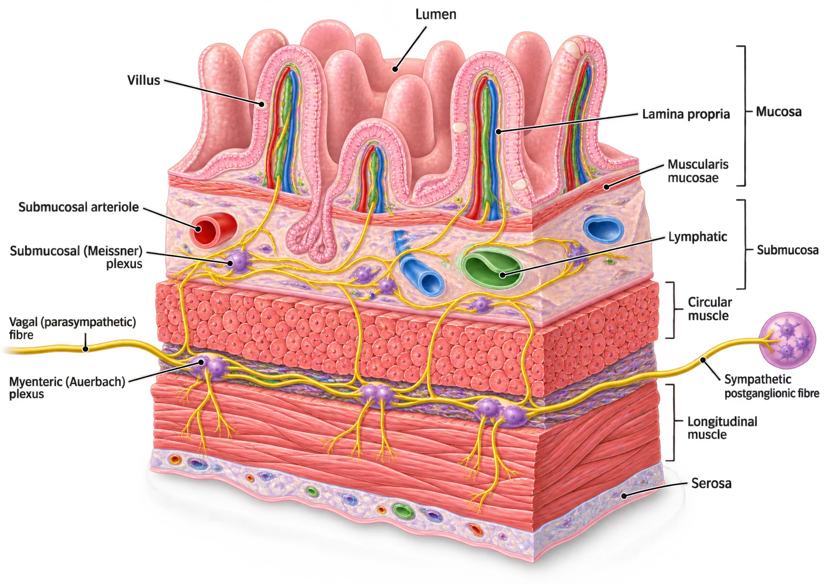
A three-dimensional cutaway of the small-intestinal wall. At the top, finger-like villi project into the lumen; each contains a central lacteal and a capillary network within the lamina propria, bounded below by the thin muscularis mucosae. Beneath the mucosa lies the submucosa, containing a submucosal arteriole, a lymphatic vessel and the submucosal (Meissner) plexus drawn as purple ganglia on a yellow nerve network. Below that is the circular muscle layer, seen end-on as bundles of cut fibres, then the myenteric (Auerbach) plexus as a sheet of ganglia between the two muscle layers, then the longitudinal muscle layer with fibres running lengthwise, and finally the serosa. A vagal parasympathetic fibre enters from the left and a sympathetic postganglionic fibre from a ganglion on the right; both terminate on the plexuses rather than on the muscle.
| Layer | What is in it | What it does |
|---|---|---|
| Serosa | — | Thin connective tissue secreting serous fluid, enclosing the viscus and reducing friction between movements. |
| Longitudinal muscle | — | Contracts to shorten the segment. |
| Myenteric (Auerbach) plexus | Between the longitudinal and circular muscle layers | Controls motility, through enteric neurones, interstitial cells of Cajal and smooth muscle. |
| Circular muscle | — | Contracts to narrow the lumen. With the longitudinal layer, produces both mixing and propulsion. |
| Submucosa | Carries the submucosal (Meissner) plexus, blood vessels and lymphatics | The submucosal plexus controls absorption, secretion and mucosal blood flow. |
| Muscularis mucosae | — | A thin smooth-muscle sheet inside the mucosa; moves the villi and keeps luminal contents from settling. |
| Lamina propria | — | Blood vessels, nerve endings, immune and inflammatory cells. In the ileum its lymphoid tissue forms Peyer's patches. |
| Epithelium | — | Where luminal contents are sensed, enzymes secreted, nutrients absorbed and waste excreted. |
Two features of the mucosa are worth separating from the rest because they are commonly confused. The muscularis mucosae is a thin sheet of smooth muscle inside the mucosa; it agitates the villi and stops luminal contents settling against the epithelium, and it is not part of the muscularis externa that produces peristalsis. The lamina propria is connective tissue carrying blood vessels, nerve endings and a standing population of immune cells; in the ileum those are organised into Peyer’s patches, which is where section 09 picks the story up.
Intrinsic control
The enteric nervous system
The enteric nervous system is the third division of the autonomic nervous system, and the only one that can complete a reflex without the central nervous system. It contains sensory neurones that respond to stretch, to luminal chemistry and to mucosal deformation; interneurones that integrate them; and motor neurones that act on smooth muscle, on secretory cells and on blood vessels. A length of intestine removed from the body and kept perfused will still generate a peristaltic wave when it is distended. That single observation is what the word “intrinsic” is doing.
| Myenteric (Auerbach) | Submucosal (Meissner) | |
|---|---|---|
| Position | Between the longitudinal and circular muscle layers | Within the submucosa |
| Controls | Motility | Secretion, absorption and mucosal blood flow |
| Cell partners | Enteric neurones, interstitial cells of Cajal, smooth muscle | Enteric neurones, secretory epithelium, submucosal arterioles |
| Extent | Continuous from oesophagus to anus | Present wherever there is a submucosa; sparse in the oesophagus |
| Loss of it | Achalasia at the lower oesophageal sphincter; aganglionic segment in Hirschsprung disease | Impaired secretion and mucosal blood flow, with less obvious clinical signature |
Extrinsic control
The extrinsic autonomic supply
| Parasympathetic | Sympathetic | |
|---|---|---|
| Origin | Medulla (vagus) and sacral cord segments S2 to S4 (pelvic nerves) | Spinal cord segments T5 to L2 |
| Distribution | Vagus to oesophagus, stomach, pancreas, small intestine and the large intestine as far as the proximal transverse colon; pelvic nerves to the remainder, sigmoid, rectum and anus | Preganglionic fibres to the coeliac, superior and inferior mesenteric ganglia, then postganglionic fibres to the gut wall |
| Preganglionic fibre length | Long | Short |
| Transmitter at the gut | Acetylcholine | Noradrenaline |
| Effect on motility and secretion | Increased | Decreased |
| Effect on sphincters | Relaxed | Contracted |
| Effect on splanchnic vessels | Little | Vasoconstriction, and venoconstriction |
| Mechanism at the plexus | Direct excitation of enteric neurones | Alpha-2 mediated inhibition of acetylcholine release from enteric neurones |
Two consequences follow that recur throughout the module. First, an intervention that reduces sympathetic outflow to the gut — a thoracic epidural, for example — increases motility and shortens the duration of postoperative ileus. Second, the same sympathetic outflow that stops the bowel also empties the splanchnic veins, which is section 08.
Endocrine control
The gastrointestinal hormones
More than fifteen types of hormone-secreting enteroendocrine cell have been identified in the mucosa of the stomach, small intestine and colon. Five of the products matter for this examination, and they fall into two structurally defined families. The gastrin family — gastrin and cholecystokinin — share the same five C-terminal amino acids, which is why the biological activity of both resides in that tail and why cholecystokinin has gastrin-like activity when it is given in large doses. The secretin family — secretin, glucagon, vasoactive intestinal peptide and gastric inhibitory peptide — share a different structure altogether.
| Hormone | Family | Cell and site | Released by | Principal actions |
|---|---|---|---|---|
| Gastrin | Gastrin | G cell, Gastric antrum; a larger form (G34) from duodenal mucosa | Vagal activity through gastrin-releasing peptide, antral distension, peptides and amino acids in the lumen. Inhibited by luminal acid through somatostatin. | Stimulates acid secretion, mostly indirectly through histamine release from enterochromaffin-like cells. Stimulates pepsinogen secretion by the chief cells. Increases gastric motility and promotes gastric emptying. Trophic to gastric, small-intestinal and colonic mucosa |
| Cholecystokinin | Gastrin | I cell, Duodenal and jejunal mucosa | Products of digestion in the duodenum: peptides, amino acids, and fatty acids of more than ten carbon atoms. | Contracts the gallbladder and relaxes the sphincter of Oddi. Stimulates pancreatic acinar enzyme secretion, and augments secretin's bicarbonate effect. Delays gastric emptying by raising pyloric tone. Trophic to the pancreas |
| Secretin | Secretin | S cell, Duodenal and upper jejunal mucosa | Acid bathing the duodenal mucosa, and the products of protein digestion. | Stimulates pancreatic and biliary duct cells to secrete a watery alkaline juice. Reduces gastric acid secretion and inhibits gastrin release. Contracts the pyloric sphincter, slowing gastric emptying |
| Gastric inhibitory peptide | Secretin | K cell, Duodenal and jejunal mucosa | Glucose and fat in the duodenum. | Stimulates insulin release at concentrations reached after an oral glucose load, which is why it is also called glucose-dependent insulinotropic peptide. Inhibits gastric acid secretion and motility, though only at doses above those reached after a meal |
| Motilin | — | M cell, Duodenal mucosa | Released cyclically during fasting; the trigger is not established. | Initiates the migrating motor complex, roughly every 90 minutes in the fasted state. Increases the force and frequency of gastric contractions. Erythromycin is a motilin agonist, which is why it is used as a prokinetic |
| Vasoactive intestinal peptide | Secretin | Enteric neurone, not an endocrine cell, Myenteric and submucosal plexuses throughout the gut | Released as a neurotransmitter, not into the circulation as a hormone. | Relaxes gastrointestinal smooth muscle, including the lower oesophageal sphincter and the pylorus. Increases intestinal secretion of water and electrolytes. One of the two non-adrenergic non-cholinergic inhibitory transmitters, with nitric oxide |
| Somatostatin | — | D cell, Gastric and duodenal mucosa, and pancreatic islets | Luminal acid, and the arrival of acidic chyme in the duodenum. | Inhibits gastrin release from G cells and acid secretion by parietal cells. Acts by paracrine diffusion rather than through the circulation |
Local control
Paracrine and immune signalling
Gastrointestinal regulation runs on three mechanisms that operate together, and the same molecule can be used in more than one of them. Endocrine signalling releases a mediator into the bloodstream to act on a distant segment or on an organ draining into it; gastrin is the standard example. Paracrine signalling releases a mediator that is too unstable to survive the circulation, so it acts only on cells in the immediate area; somatostatin from the D cell, diffusing onto the neighbouring G cell and parietal cell, is the standard example, and so is histamine from the enterochromaffin-like cell. Neurocrine signalling releases a transmitter from a nerve terminal; vasoactive intestinal peptide and nitric oxide from the inhibitory enteric neurones are the standard examples, and they are the pair that relaxes every sphincter in the tract.
Circulation
The splanchnic circulation
The splanchnic circulation is unlike any other regional bed in one specific respect: its venous outflow does not return to the heart. It returns to another organ. Blood from the stomach, spleen, pancreas, small intestine and colon drains into the portal vein and perfuses the hepatic sinusoids before entering the hepatic veins, the inferior vena cava and the right atrium. That single arrangement explains first-pass metabolism, portal hypertension, the site of a gastrointestinal haemorrhage’s effect on ammonia, and why the liver is the first organ to see anything absorbed from the gut.
| Vessel | Fasting flow | Territory |
|---|---|---|
| Coeliac artery | 700 mL/min | Stomach, spleen, pancreas, liver, proximal duodenum |
| Superior mesenteric artery | 700 mL/min | Distal duodenum to the proximal transverse colon |
| Inferior mesenteric artery | 400 mL/min | Distal transverse colon to the upper rectum |
Those three sum to 1800 mL/min, and the viscera together with the liver receive about 30% of the cardiac output at rest. Feeding raises it substantially; vigorous exercise, haemorrhage and any other state of high sympathetic tone reduce it sharply, and the splanchnic bed is one of the first to be sacrificed. That vulnerability is the physiological basis of non-occlusive mesenteric ischaemia in a patient on high-dose vasoconstrictors, and of the mucosal injury that follows a prolonged period of low cardiac output.
The splanchnic bed at rest, and under sympathetic stimulation
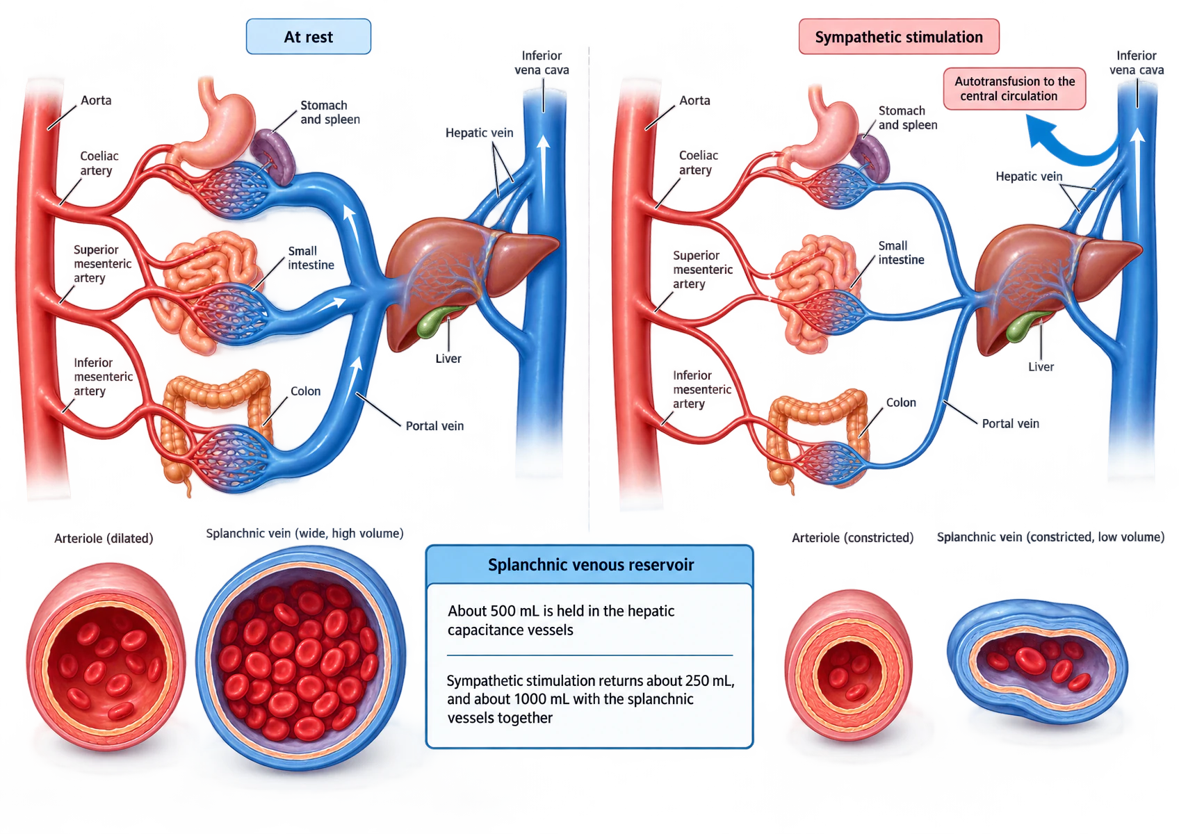
Two side-by-side panels. In the left panel, labelled "At rest", the abdominal aorta gives off the coeliac, superior mesenteric and inferior mesenteric arteries, which supply capillary networks in the stomach and spleen, the small intestine and the colon. All three networks drain into a wide portal vein, which enters the liver; hepatic veins leave the liver for the inferior vena cava. The right panel, labelled "Sympathetic stimulation", shows the same anatomy with narrowed arteries and a narrowed portal vein, and a broad arrow marked "autotransfusion to the central circulation" running from the hepatic veins into the inferior vena cava. Along the foot of the figure, four vessel cross-sections compare a dilated arteriole and a wide splanchnic vein packed with red cells at rest against a constricted arteriole and a collapsed, nearly empty vein under stimulation. A panel between them states that about 500 mL is held in the hepatic capacitance vessels, that sympathetic stimulation returns about 250 mL, and about 1000 mL with the splanchnic vessels together.
Circulation
The gut as a blood reservoir
The splanchnic veins, the portal venules and the hepatic veins are thin-walled, compliant and densely innervated by sympathetic fibres. At rest they hold a large volume at low pressure. When sympathetic tone rises — on standing, on exercise, on haemorrhage — those vessels constrict, and the volume they were holding is returned to the central circulation. The effect is an autotransfusion, and it happens before any of the slower compensations for hypovolaemia have begun.
Defence
Barrier function
The gastrointestinal epithelium is a single cell thick over an absorptive area of hundreds of square metres, and on its luminal side sits a bacterial population outnumbering the body’s own cells. The barrier that separates them has four components, and each fails in a way that shows up in the perioperative period.
| Component | What it is | What defeats it |
|---|---|---|
| Physical | A continuous epithelium sealed by tight junctions, over an unstirred mucus layer secreted by goblet cells, with an antimicrobial peptide gradient within it | Hypoperfusion, which kills the villous tip first because its counter-current capillary arrangement makes it the most vulnerable point in the mucosa |
| Chemical | Gastric acid, bile acids, pancreatic proteases and lysozyme, each of which kills or digests organisms in transit | Acid suppression, which raises gastric pH and permits bacterial colonisation of the stomach |
| Immunological | Gut-associated lymphoid tissue: Peyer's patches in the ileum, plasma cells in the lamina propria secreting IgA, intraepithelial lymphocytes | Malnutrition, and the loss of enteral feeding, which reduces IgA secretion |
| Microbiological | The resident flora, which occupies niches and produces short-chain fatty acids that nourish the colonocyte | Broad-spectrum antibiotics, and the mechanical loss of the migrating motor complex, which normally sweeps bacteria distally |
Secretory immunoglobulin A is the specific immunological point worth carrying. It is the most abundantly produced immunoglobulin in the body, it is secreted as a dimer with a secretory component that protects it from luminal proteases, and it works by immune exclusion — binding organisms and preventing their adherence rather than opsonising them for destruction. That is why it does not fix complement and why it does not provoke an inflammatory response every time it does its job.
Consolidation
The lesson in one paragraph
The gastrointestinal tract is a nine-metre tube built on one plan of wall: serosa, longitudinal muscle, circular muscle, submucosa and mucosa, with the mucosa itself divided into muscularis mucosae, lamina propria and epithelium. Two nerve plexuses sit at fixed addresses within that plan, and their addresses give their functions — the myenteric plexus between the two muscle layers controls motility through enteric neurones, the pacemaking interstitial cells of Cajal and smooth muscle, and the submucosal plexus within the submucosa controls secretion, absorption and mucosal blood flow. Together they constitute the enteric nervous system, a complete reflex arc that runs peristalsis with every extrinsic nerve cut; the extrinsic supply modulates it rather than driving it. Parasympathetic fibres, vagal to the proximal transverse colon and pelvic beyond it, are excitatory through acetylcholine; sympathetic fibres from T5 to L2 are inhibitory through noradrenaline at alpha-2 receptors on enteric neurones, and at the same time contract the sphincters and constrict the splanchnic vessels, so wall tone rises while transit falls. Endocrine control runs on two structural families: gastrin and cholecystokinin, which share five C-terminal amino acids, and secretin, glucagon, vasoactive intestinal peptide and gastric inhibitory peptide. Gastrin and motilin prepare and sweep the stomach and raise sphincter tone; secretin, cholecystokinin and gastric inhibitory peptide are released by what has already left the stomach and slow it down. Local control adds paracrine signalling, of which somatostatin and histamine are the examples, and neurocrine signalling, of which nitric oxide and vasoactive intestinal peptide are the inhibitory pair. The circulation is arranged as three arterial trunks supplying three capillary beds in parallel — coeliac 700, superior mesenteric 700 and inferior mesenteric 400 mL/min, about 30% of the cardiac output — all of which then drain in series through a second capillary bed in the liver before reaching the heart. Those veins are the body’s largest blood reservoir: about 500 mL in the hepatic capacitance vessels, about 250 mL of it returned on sympathetic stimulation, and about 1000 mL from the splanchnic bed as a whole, which is why abolishing sympathetic tone at induction unmasks a hypovolaemia that had been concealed. Guarding all of it is a barrier with physical, chemical, immunological and microbiological components, of which the villous tip is the most vulnerable to hypoperfusion and secretory IgA the most abundant defence.