Fluid mosaic model
The membrane is a fluid lipid bilayer studded with functional proteins
What you should already have
About 40 minutes
Plus the time it takes to redraw this lesson’s figures from memory, which is the fastest way to find out what you have not understood.
Where this shows up
This is where pharmacokinetics starts. Whether a drug crosses a membrane at all, by which route, and whether that route can saturate or be competed for, are all decided by the mechanisms here. It is also where the fluid prescription in front of you is decided: osmolality and tonicity are not the same quantity, and only one of them predicts what happens to the cell.
Learning outcomes
By the end of this lesson you should be able to:
- Describe the fluid mosaic membrane and explain why selective permeability does not mean the membrane is closed.
- State why lipid solubility is the single best predictor of simple permeability, and what that predicts for a drug.
- Explain diffusion as random movement with a directional net result, and give the factors that set its rate.
- Classify GLUT-mediated glucose transport correctly as facilitated diffusion, and say what distinguishes it from an active process.
- Distinguish primary pumps, which create gradients, from secondary transport, which spends them.
- Describe vesicular transport, and say what cargo requires it.
- Separate osmolality from tonicity and predict the cell-volume consequence of a given solution.
Together these settle one syllabus objective: Membrane transport, osmosis and tonicity. Tick it on the Physiology objective list once you can do all of the above without notes.
The plasma membrane — fluid mosaic model
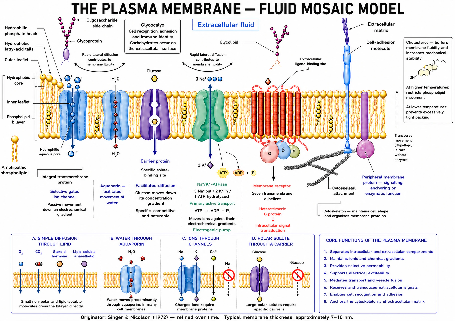
Phospholipids are amphipathic: a hydrophilic phosphate head faces water on either side, while two hydrophobic fatty-acid tails pack together to form the bilayer core. Each leaflet is a mirror image of the other, so the bilayer is a two-layer sandwich with hydrophilic surfaces and a hydrophobic centre — the arrangement that makes the membrane a barrier to water-soluble and charged solutes at the same time as it stays fluid enough for proteins to move within it.
Composition: phospholipids, cholesterol, proteins and carbohydrates
| Component | Location | Structural role | Why it matters physiologically |
|---|---|---|---|
| Phospholipids | Both leaflets | Form the bilayer itself; tail saturation sets packing density | The barrier to water-soluble and charged solutes |
| Cholesterol | Intercalated among the tails, hydroxyl at the head region | Modulates fluidity and mechanical stability (below) | Keeps the membrane functional across a temperature range |
| Integral (transmembrane) proteins | Span the bilayer | Channels, carriers, pumps, receptors | Do the work a lipid bilayer alone cannot |
| Peripheral proteins | Attached to one face only, usually cytoplasmic | Anchoring, signalling scaffolds, cytoskeletal linkage | Shape, mechanical support, localisation of signalling |
| Glycoproteins and glycolipids | Carbohydrate chains project into extracellular fluid only | Cell recognition, adhesion, blood-group and immune identity | Restricted sidedness (glycocalyx) makes the membrane asymmetric, not just layered |
Cholesterol’s effect on fluidity is temperature-dependent, and the direction matters. Above the membrane’s phase-transition temperature, phospholipid tails are relatively disordered and mobile; cholesterol’s rigid steroid ring system wedges between them, restricts their movement and reduces fluidity. Below the phase-transition temperature, tails would otherwise pack tightly into an ordered, gel-like state; cholesterol disrupts that close packing and increases fluidity, preventing the membrane from becoming too rigid. The net effect is that cholesterol narrows the temperature range over which the membrane’s fluidity changes sharply — it acts as a buffer, keeping the bilayer in a workable intermediate state rather than letting it swing between a rigid gel and an overly fluid liquid as temperature changes. This is why cholesterol content, not just temperature, has to be considered whenever membrane function is discussed across a temperature range, from deliberate intraoperative hypothermia to fever.
What crosses, and why
Lipid solubility is the single best predictor of simple permeability
Small, uncharged, lipid-soluble molecules (O₂, CO₂, volatile anaesthetic agents, steroid hormones) cross the bilayer directly and rapidly. Water crosses faster than its lipid solubility alone predicts, mainly through aquaporin channels rather than through the lipid itself. Ions and polar solutes (Na⁺, K⁺, Ca²⁺, glucose, amino acids) are essentially excluded from the hydrophobic core and depend entirely on the transport proteins covered in the rest of this lesson.
Anaesthetic relevance — the Meyer–Overton correlation and the theories of anaesthesia
The potency of an inhalational anaesthetic correlates closely with its solubility in olive oil (a lipid surrogate) across an enormous range of chemically unrelated agents — the Meyer–Overton correlation. Historically this was read as evidence that anaesthetics act by dissolving in the bulk lipid bilayer itself and disordering it (the lipid or “membrane expansion” theories of anaesthesia). The correlation is real and durable, but the mechanism it was thought to support has not held up: bulk lipid disruption of the kind these theories proposed does not reproduce the anaesthetic state.
Current evidence instead points to direct, saturable binding at discrete amphiphilic cavities within specific membrane proteins — a protein-target theory that keeps the same correlation (a hydrophobic binding pocket still favours a lipid-soluble drug) without needing bulk lipid disruption. Volatile agents potentiate inhibitory ligand-gated channels (GABA-A, glycine) and inhibit excitatory glutamate channels (NMDA, AMPA, among others); no single protein target accounts for the whole anaesthetic state, and different components of anaesthesia (immobility, hypnosis, amnesia) appear to depend on different sites. The lipid-solubility correlation and the protein-binding mechanism are not competing facts — the correlation is the observation; the protein-binding theory is today’s best explanation for it.
Passive movement
Diffusion is random movement with a directional net result
| Symbol | Meaning | SI units | Effect on diffusion |
|---|---|---|---|
| ṅ | Amount crossing per unit time | mol s⁻¹ | The result |
| P | Permeability; incorporates partition coefficient, diffusion coefficient and membrane thickness | m s⁻¹ | Greater P increases flux |
| A | Surface area available for exchange | m² | Greater area increases flux |
| C₁ − C₂ | Concentration difference across the membrane | mol m⁻³ | Greater gradient increases flux |
Equivalently, permeability can be expressed as P = K·D/Δx, where K is the lipid:water partition coefficient, D is the diffusion coefficient within the membrane and Δx is membrane thickness: a higher partition coefficient or diffusion coefficient increases diffusion, whereas a thicker membrane reduces it. Small molecules and low-viscosity media generally have higher diffusion coefficients. Charge and ionisation reduce simple diffusion through lipid, because a charged species partitions poorly into a hydrophobic core.
Worked example
If P = 2 × 10⁻⁵ m s⁻¹, A = 1 × 10⁻⁴ m² and C₁ − C₂ = 10 mol m⁻³, then ṅ = P × A × (C₁ − C₂) = (2 × 10⁻⁵)(1 × 10⁻⁴)(10) = 2 × 10⁻⁸ mol s⁻¹. Sanity check: doubling area doubles flux; doubling membrane thickness halves P and therefore halves flux — both match the direct/inverse relationships in the equation.
Predict what happens if: a burn or inflammation doubles capillary surface area available for exchange, with everything else unchanged — flux roughly doubles, since ṅ is directly proportional to A.
Protein-mediated, still passive
Channels and carriers make specific solutes cross faster, without spending energy
| Protein | Physical mechanism | Key property | Rate behaviour | Example |
|---|---|---|---|---|
| Ion channel | Water-filled selective pore | Open probability/gating | High conductance when open | Voltage-gated Na⁺ channel, aquaporin |
| Carrier | Binds solute and alternately exposes its binding site | Specificity, competition, saturation | Finite transport maximum (Tmax) | Glucose transporter (GLUT) |
| Pump | Carrier coupled directly to a chemical energy source | Specificity and saturation | Moves solute against its gradient | Na⁺/K⁺-ATPase |
| Symporter | Couples two solutes moving in the same direction | Uses energy stored in a gradient | Secondary active | Na⁺–glucose cotransporter (SGLT) |
| Antiporter | Couples two solutes moving in opposite directions | Uses energy stored in a gradient | Secondary active | Na⁺/Ca²⁺ exchanger |
Facilitated diffusion is passive despite requiring a carrier: glucose moves down its electrochemical gradient through GLUT transporters without any ATP being spent. Because the number of carrier molecules is finite, facilitated diffusion shares three properties with every other carrier-mediated process in this lesson (including the pumps in the next section):
- Specificity — a carrier recognises a defined solute or closely related family of solutes, unlike the bilayer itself, which discriminates only by lipid solubility, size and charge.
- Saturation — rate rises with concentration only until every carrier is occupied, at which point the rate plateaus at a transport maximum (Tmax), no matter how much further the gradient steepens.
- Competition — a structurally similar molecule can occupy the same binding site and reduce the transport rate of the solute you are interested in.
Facilitated diffusion saturates; simple diffusion does not
A finite number of carrier proteins gives facilitated diffusion a transport maximum; simple diffusion through the lipid bilayer keeps rising as concentration rises. Axis values are illustrative (arbitrary units), not measured data.
Predict what happens if: extracellular glucose concentration is pushed far above the physiological range — facilitated glucose entry via GLUT approaches its transport maximum and stops rising in proportion, unlike simple diffusion of a lipid-soluble gas, which keeps rising linearly with the gradient.
Spending energy to move uphill
Primary pumps create gradients directly; secondary transport spends them
The sodium–potassium adenosine triphosphatase (Na⁺/K⁺-ATPase) moves three sodium ions out and two potassium ions in for each ATP hydrolysed. It is electrogenic (the unequal 3:2 exchange contributes a small direct current), but its dominant physiological importance is maintaining the sodium and potassium concentration gradients that everything else in this module depends on: cell-volume regulation, the resting membrane potential and, through secondary active transport, the uphill movement of glucose, amino acids and calcium.
Sarco/endoplasmic reticulum Ca²⁺-ATPase (SERCA) uses ATP directly to sequester calcium into the endoplasmic or sarcoplasmic reticulum; the plasma-membrane Ca²⁺-ATPase extrudes calcium from the cell by the same primary-active mechanism. Both keep resting cytosolic calcium roughly four orders of magnitude below extracellular calcium — the steep gradient that makes calcium useful as a fast intracellular signal (developed further in the signalling lesson).
Secondary active transport spends, rather than creates, a gradient. The sodium-glucose cotransporter (SGLT) uses the inward electrochemical pull on Na⁺ (built by the Na⁺/K⁺-ATPase) to drag glucose uphill into the cell — a symporter, because both solutes move the same direction. The Na⁺/Ca²⁺ exchanger uses the same stored sodium gradient to extrude calcium — an antiporter, because the two solutes move in opposite directions. Neither hydrolyses ATP itself; both stop working if the Na⁺/K⁺-ATPase is poisoned (for example by a cardiac glycoside indirectly raising intracellular Na⁺), which is why “active transport” is defined by the direction of net movement against a gradient, not by which specific step consumes ATP.
All seven routes across the membrane, compared
Every mechanism in this lesson answers the same four questions differently: what carries the solute, which way it moves relative to its gradient, whether it saturates, and where the energy comes from. Read across a row before comparing rows — the pattern that matters for the exam is which properties travel together (saturation, specificity and competition are a package; simple diffusion and filtration are the only two that have none of them).
| Mechanism | Carrier | Direction relative to gradient | Saturates | Energy source | Example |
|---|---|---|---|---|---|
| Simple diffusion | None | Down concentration gradient | No | No | O₂, CO₂, volatile anaesthetic agents, other lipid-soluble molecules |
| Channel-mediated diffusion | Ion channel | Down electrochemical gradient | Yes (gating), not saturation in the Michaelis–Menten sense | No | K⁺ leak channels, ligand-gated Na⁺ channels |
| Facilitated (carrier-mediated) diffusion | Carrier | Down electrochemical gradient | Yes — saturates at Tmax | No | GLUT-mediated glucose entry |
| Primary active transport | ATPase pump | Against electrochemical gradient | Yes — saturates | Direct (ATP hydrolysis) | Na⁺/K⁺-ATPase; sarco/endoplasmic reticulum Ca²⁺-ATPase (SERCA) |
| Secondary active transport | Cotransporter / exchanger | One solute downhill drives another uphill | Yes — saturates | Indirect (a pre-existing gradient) | Na⁺-glucose cotransporter (SGLT); Na⁺/Ca²⁺ exchange |
| Filtration / bulk flow | None (pressure-driven) | Down a hydrostatic or osmotic pressure gradient | No | No | Capillary fluid movement (see Starling forces, fluids lesson) |
| Vesicular transport | Vesicle formation and membrane fusion | Bulk transfer of cargo too large for a channel or carrier | No | Cellular ATP-dependent machinery | Receptor-mediated endocytosis; regulated exocytosis |
Bulk transport
Vesicular transport moves cargo too large for a channel or carrier
| Process | Direction | Mechanism | Anaesthesia-relevant example |
|---|---|---|---|
| Phagocytosis | Into the cell | Large particles engulfed by membrane extensions | Neutrophil/macrophage clearance of pathogens and debris |
| Pinocytosis / fluid-phase endocytosis | Into the cell | Non-specific uptake of extracellular fluid and solutes | Bulk membrane and protein turnover |
| Receptor-mediated endocytosis | Into the cell | Ligand-receptor complex internalised via clathrin-coated pits | Receptor desensitisation and internalisation after prolonged agonist exposure — one mechanism of tolerance |
| Regulated (Ca²⁺-triggered) exocytosis | Out of the cell | Vesicle fuses with the membrane on a rise in cytosolic Ca²⁺ | Neurotransmitter release at the neuromuscular junction and central synapses |
| Constitutive exocytosis | Out of the cell | Continuous, unregulated vesicle fusion | Insertion of newly synthesised membrane proteins and receptors |
Vesicular transport shares one feature with primary active transport and none with simple diffusion: it is an energy-dependent, cellular process (vesicle budding, cytoskeletal transport and fusion machinery all consume ATP or GTP), and — unlike every other process in this lesson — it moves bulk cargo rather than individual solute molecules one at a time.
Water movement
Osmolality describes particles; tonicity predicts cell volume
Tonicity predicts the cell-volume response
Water moves toward the side with higher effective osmotic pressure. Labels assume a membrane that is poorly permeable to the effective extracellular solute.
Cell swells
Volume unchanged
Cell shrinks
| Symbol | Meaning | Units |
|---|---|---|
| π | Effective (colloid or crystalloid) osmotic pressure | Pa (or mmHg by convention for oncotic pressure) |
| σ | Reflection coefficient: 0 = solute freely crosses the membrane, 1 = solute is completely impermeant | Dimensionless, 0–1 |
| i | Van 't Hoff (dissociation) factor — particles produced per dissolved molecule | Dimensionless |
| C | Solute concentration | mol m⁻³ |
| R | Universal gas constant | J mol⁻¹ K⁻¹ |
| T | Absolute temperature | K |
| Term | Definition | Why it matters |
|---|---|---|
| Osmolarity | Osmoles of solute per litre of solution | Volume-dependent description used clinically as an estimate |
| Osmolality | Osmoles of solute per kilogram of solvent | Mass-dependent — the value a laboratory freezing-point osmometer actually measures |
| Osmotic pressure | Pressure required to stop net osmotic water flow | Rises with effective (not total) particle concentration |
| Reflection coefficient, σ | How completely a membrane excludes a solute | A non-penetrating solute (σ near 1) exerts its full osmotic effect; a freely penetrating one (σ near 0) exerts almost none, however concentrated |
| Tonicity | A solution’s net effect on cell volume | Depends on effective osmoles only — a property of the solute-membrane pair, not of the solution alone |
Urea contributes to measured osmolality but exerts little sustained tonic effect on most cell membranes, because it eventually equilibrates across them (a low reflection coefficient) — a rise in plasma urea alone does not sustainedly shrink cells the way an equivalent rise in an impermeant solute would. Sodium salts, largely excluded from cells by the Na⁺/K⁺-ATPase, remain effective extracellular osmoles and do determine sustained cell volume. Acute water movement (the immediate osmotic response) and the eventual steady state (once any permeant solute has redistributed) can therefore differ substantially for the same starting osmolality.
Worked example — osmolarity vs osmolality, and why the distinction matters
A solution contains 150 mmol L⁻¹ NaCl (i = 2, fully dissociating) and 5 mmol L⁻¹ glucose (i = 1). Osmolarity = (150 × 2) + (5 × 1) = 305 mOsm L⁻¹. If this same solution is prepared in 1 L of water whose mass is 0.994 kg once the solutes are dissolved (typical for a dilute aqueous solution), osmolality = 305 mmol ÷ 0.994 kg ≈ 307 mOsm kg⁻¹ — numerically close but not identical, because osmolarity is volume-based and osmolality is mass-based. The gap widens as solute concentration rises (more dissolved mass per litre of solution), which is why concentrated protein-rich or lipid-rich plasma samples can show a measurable osmolar gap between calculated osmolarity and measured osmolality.
Hypotonic extracellular fluid drives net water entry and cell swelling; in the brain, this can worsen cerebral oedema — one reason hypotonic intravenous fluids are avoided in neurological injury. Hypertonic extracellular fluid drives net water loss and cell shrinkage, the basis for using hypertonic saline or mannitol (a non-penetrating solute at the blood-brain barrier) to draw water out of brain tissue and reduce intracranial pressure.
Take-home points for this lesson. The bilayer’s hydrophobic core is the barrier; lipid solubility predicts what crosses it unassisted, and that same relationship (the Meyer–Overton correlation) is the historical basis for the lipid theories of anaesthesia, now superseded by protein-target theories that still respect the correlation. Every carrier-mediated process — facilitated diffusion, primary active transport, secondary active transport — is specific, saturable and competitively inhibited; only simple diffusion and filtration are not. Active transport is defined by movement against a gradient, not by which step spends the ATP. Osmolarity counts particles; tonicity depends on the reflection coefficient of each particle at that particular membrane.