What you should already have
About 80 minutes
Plus the time it takes to redraw this lesson’s figures from memory, which is the fastest way to find out what you have not understood.
Where this shows up
The functions of the liver are asked more often than anything else in this section, and the commonest way of losing marks is to know them all and present them as an unordered list. This lesson gives the headings first and hangs everything off them, because a structure you can reproduce under pressure is worth more than a longer set of facts you cannot organise.
Learning outcomes
By the end of this lesson you should be able to:
- Classify the functions of the liver under headings that survive an examination answer, and give the consequence of losing each.
- Describe the liver's handling of glucose in the fed and the fasting state, and state the size of the glycogen store.
- Describe deamination, transamination and the urea cycle, and explain why ammonia rises in liver failure and why the concentration does not predict encephalopathy.
- State the liver's roles in lipid metabolism, including ketogenesis and lipoprotein assembly.
- Name the coagulation factors the liver does and does not synthesise, and separate the vitamin K-dependent factors from the rest.
- Describe bile: its volume, its constituents, how bile acids are made and how the enterohepatic circulation recycles them.
- Trace bilirubin from senescent red cell to urine and stool, and use conjugated against unconjugated hyperbilirubinaemia to classify jaundice.
- State the liver's storage, immunological and protective roles, and its part in acid-base balance through the urea cycle and glutamine synthesis.
- Interpret a standard liver panel: what each test measures, what a change means, and the limitation of each.
- Define acute liver failure and describe the metabolic, synthetic and neurological consequences of it.
Together these settle one syllabus objective: The metabolic, synthetic, storage, protective and excretory functions of the liver, their assessment, and acute liver failure. Tick it on the Physiology objective list once you can do all of the above without notes.
Orientation
Rapid review
- Six headings. Metabolic, synthetic, storage, excretory and exocrine, protective and immunological, endocrine and regulatory. Learn the headings first; the content hangs off them and can be rebuilt.
- The liver makes everything except immunoglobulins, factor VIII and tissue factor. That is a shorter and more useful statement than any list of what it does make.
- Prothrombin time moves in a day, albumin in a month. Factor VII’s half-life is about six hours and albumin’s about three weeks, which is why a normal albumin does not exclude acute liver failure.
- The urea cycle consumes bicarbonate. In acidosis the liver switches from ureagenesis to glutamine synthesis, sparing bicarbonate and handing the ammonium to the kidney. This is the liver’s role in acid-base balance and it is usually missed.
- Jaundice classifies on the conjugated fraction and on urinary bilirubin. Only conjugated bilirubin is water-soluble, so only it appears in urine.
- Acute liver failure is a definition with four parts: encephalopathy, coagulopathy with an INR of 1.5 or more, no previous liver disease, and an illness of under 26 weeks. Cerebral oedema is what kills.
Metabolic
Carbohydrate metabolism
| Process | When | What happens | Driven by |
|---|---|---|---|
| Glycolysis | Continuously | Glucose to pyruvate, for the liver's own energy. Predominantly a zone 3 process | — |
| Glycogenesis | Fed | Excess glucose is polymerised to glycogen. Up to about 100 g is stored this way | Insulin |
| Lipogenesis | Fed, once glycogen stores are full | Further excess glucose is converted to triglyceride and exported as VLDL | Insulin |
| Glycogenolysis | Early fasting | Glycogen is broken back down to glucose and released. The first-line defence, and exhausted within about 24 hours | Glucagon, and adrenaline |
| Gluconeogenesis | Later fasting | Glucose is synthesised from lactate, pyruvate, glycerol and glucogenic amino acids. Predominantly a zone 1 process | Glucagon, cortisol |
The Cori cycle is worth naming here because it reappears in section 10. Lactate produced by anaerobic glycolysis in muscle and red cells travels to the liver, is converted back to glucose by gluconeogenesis, and returns. The liver is therefore the principal site of lactate disposal in the body, which is why hepatic failure and lactic acidosis go together and why a rising lactate in a patient with a normal cardiac output should raise the question of hepatic clearance rather than only of tissue hypoxia.
Metabolic
Protein metabolism, urea and ammonia
- Transamination. An amino group is moved from an amino acid to a keto acid, making a different amino acid. This is how the eleven non-essential amino acids are made, and it is the reaction the aminotransferases catalyse — the same enzymes whose leakage is measured as ALT and AST.
- Deamination. The amino group is removed altogether, leaving a carbon skeleton that enters the citric acid cycle or gluconeogenesis, and releasing ammonia.
- The urea cycle. Ammonia is toxic, particularly to the brain, and is converted to urea, which is not. The cycle spans the mitochondrion and the cytosol of the periportal hepatocyte, and the urea is excreted by the kidney.
- Glutamine synthesis. A second, high-affinity route, in the perivenous cells. Ammonia that escaped the periportal urea cycle is scavenged here and exported as glutamine.
Ammonia arises from two sources: deamination in the liver itself, and colonic bacterial breakdown of protein and urea, which reaches the liver in the portal blood. That second source is why a gastrointestinal bleed precipitates encephalopathy — blood in the lumen is a protein meal — and why lactulose and rifaximin work, one by acidifying the colon so that ammonia is trapped as ammonium and not absorbed, the other by reducing the bacteria producing it.
Metabolic
Fat metabolism
| Role | What happens | Why it matters |
|---|---|---|
| Beta-oxidation | Free fatty acids are oxidised in the hepatocyte mitochondria to acetyl-CoA, releasing energy. Predominantly a zone 1 process | The liver's own principal fuel in the fasted state |
| Ketogenesis | When acetyl-CoA exceeds what the citric acid cycle can take — in starvation, or in uncontrolled diabetes — it is converted to acetoacetate and beta-hydroxybutyrate. A zone 3 process | The liver makes ketone bodies and cannot use them. They are a fuel for the brain and other tissues, and the liver is the only organ that produces them |
| Lipogenesis | Excess carbohydrate and protein are converted to fatty acids and esterified to triglyceride | How a surplus of any macronutrient ends up as fat |
| Cholesterol synthesis | The liver is the principal site, and cholesterol is the precursor of the bile acids and of the steroid hormones | Also the route of cholesterol elimination, through bile — section 06 |
| Lipoprotein assembly | Apolipoproteins are synthesised here and used to package lipid as VLDL, and to receive it as LDL and HDL | Lipid cannot travel in plasma unpackaged, so hepatic synthetic failure disturbs the whole lipid profile |
Synthetic
Synthetic function and coagulation
| Group | Members | Note |
|---|---|---|
| Vitamin K-dependent factors | II, VII, IX and X, with protein C and protein S | Vitamin K is the cofactor for the gamma-carboxylation that lets these bind calcium and attach to phospholipid surfaces. Warfarin inhibits that carboxylation, which is why its effect looks identical to vitamin K deficiency |
| Other coagulation factors | I (fibrinogen), V, XI, XIII | Not vitamin K-dependent, so unaffected by warfarin or by biliary obstruction |
| Anticoagulant and fibrinolytic proteins | Antithrombin III, protein C, protein S, plasminogen, alpha-2 antiplasmin | These fall alongside the procoagulants in liver disease, which is why a prolonged INR there does not predict bleeding the way it does in warfarin therapy |
| Albumin | Over half of total plasma protein | Carries drugs, bilirubin, hormones and free fatty acids, and provides most of the plasma oncotic pressure |
| Transport proteins | Transferrin, caeruloplasmin, haptoglobin, thyroxine-binding globulin, sex hormone-binding globulin, alpha-1 acid glycoprotein | Alpha-1 acid glycoprotein binds basic drugs and is an acute-phase reactant, so binding changes in illness |
| Acute-phase and protective proteins | C-reactive protein, complement, alpha-1 antitrypsin | Section 09 |
Excretory
Bile and the enterohepatic circulation
Bile, from hepatocyte to terminal ileum and back
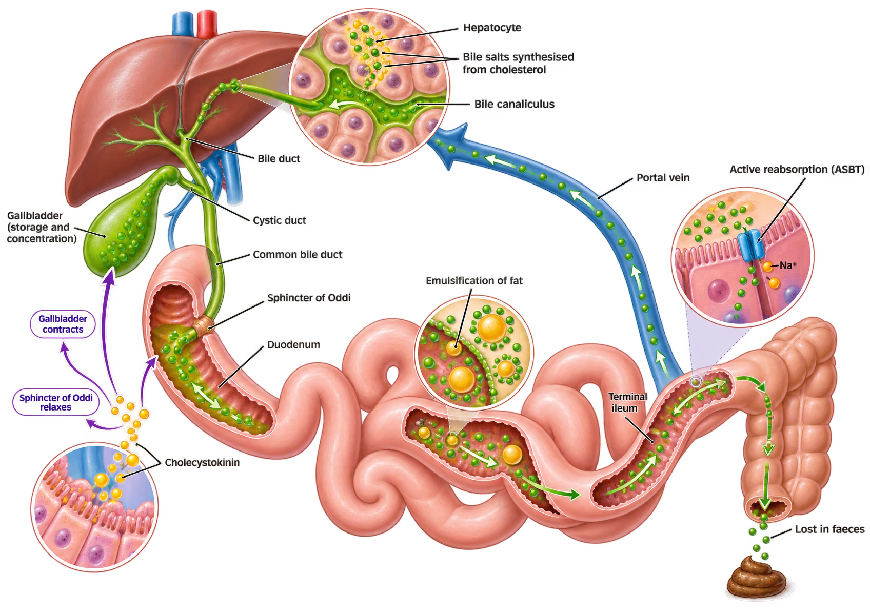
A schematic of bile formation and the enterohepatic circulation. Top left, the liver, with an inset circle magnifying hepatocytes secreting green bile salts, synthesised from cholesterol, into a bile canaliculus between them. Bile drains through intrahepatic bile ducts to the common hepatic duct, and by the cystic duct to the gallbladder, labelled as the site of storage and concentration. The common bile duct carries bile to the duodenum through the sphincter of Oddi. An inset at lower left shows duodenal mucosal cells releasing cholecystokinin, with arrows indicating that it contracts the gallbladder and relaxes the sphincter of Oddi. In the small intestine an inset shows emulsification, with bile salts surrounding large yellow fat droplets and breaking them into smaller ones. At the terminal ileum a further inset shows active reabsorption of bile salts by the ASBT transporter, coupled to sodium. A blue portal vein carries the reabsorbed salts back to the liver. At the far right a small quantity of bile salts passes into the colon and is lost in faeces.
| Detail | |
|---|---|
| Volume | 400 to 1000 mL a day — the sources differ, one giving 400 to 600 and another about 1000. Concentrated about fivefold in the gallbladder |
| Composition | 95% water, with bile acids, phospholipids, cholesterol, bilirubin and electrolytes |
| Primary bile acids | Cholic acid and chenodeoxycholic acid, synthesised from cholesterol, then conjugated with glycine or taurine to reduce toxicity and increase solubility |
| Digestive function | Bile salts are amphipathic and emulsify dietary fat, and form the mixed micelles without which lipase cannot act and the fat-soluble vitamins cannot be absorbed |
| Excretory function | The route of elimination for compounds above about 300 to 500 daltons, which the kidney does not filter well: bilirubin, cholesterol, steroid hormones, drugs and their conjugates |
| Released by | Cholecystokinin, from duodenal fat and protein products, which contracts the gallbladder and relaxes the sphincter of Oddi |
Excretory
Bilirubin
Bilirubin, from senescent red cell to urine and stool
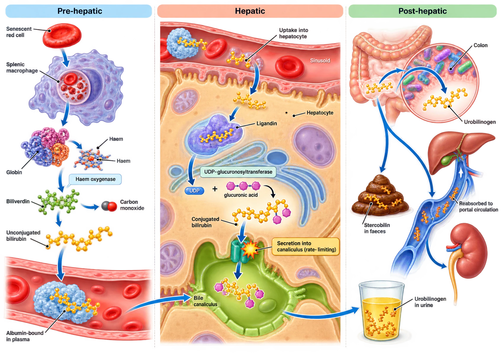
Two typographical defects in the supplied artwork, neither affecting the pathway. “Haem” is labelled twice on the same porphyrin molecule, and the secretion box reads “rate- limiting” with a stray space. One omission rather than an error: iron recycling to transferrin is not drawn, although globin is.
Three vertical panels. The pre-hepatic panel shows a senescent red cell taken up by a splenic macrophage; haemoglobin separates into globin and haem; haem oxygenase converts the porphyrin ring to green biliverdin, releasing carbon monoxide; biliverdin is reduced to yellow unconjugated bilirubin, which is shown bound to albumin within a blood vessel. The hepatic panel shows a hepatocyte beside a sinusoid: bilirubin is taken up from the sinusoid, bound to ligandin, conjugated with glucuronic acid by UDP-glucuronosyltransferase, and the conjugated product transported into the bile canaliculus, this last step marked as rate-limiting. The post-hepatic panel shows conjugated bilirubin entering the colon where bacteria convert it to urobilinogen, which has three fates: oxidation to stercobilin and loss in faeces, reabsorption into the portal circulation and return to the liver, and renal excretion as urobilinogen in urine.
- Production. Senescent red cells are phagocytosed by macrophages of the spleen, liver and bone marrow. Haem oxygenase opens the porphyrin ring to biliverdin, releasing carbon monoxide and iron; the iron returns to transferrin, and biliverdin reductase makes bilirubin.
- Transport. Unconjugated bilirubin is lipid-soluble and not water-soluble, so it travels tightly bound to albumin and is not filtered at the glomerulus. This one property does most of the diagnostic work below.
- Uptake and conjugation. The hepatocyte takes it up, binds it to ligandin, and conjugates it with glucuronic acid using UDP-glucuronosyltransferase, making it water-soluble.
- Secretion. Conjugated bilirubin is actively transported into the canaliculus. This is the rate-limiting step of the whole pathway, which is why hepatocellular injury tends to raise the conjugated fraction.
- Intestinal fate. Colonic bacteria deconjugate and reduce it to urobilinogen. Some is oxidised to stercobilin, which colours the stool; some is reabsorbed and re-excreted; a fraction escapes the liver and is excreted in urine as urobilinogen, which colours it.
| Pre-hepatic | Hepatocellular | Post-hepatic | |
|---|---|---|---|
| Cause | Excessive production, or failure of uptake or conjugation | Hepatocyte injury and disruption of the biliary tree | Obstruction to the flow of bile |
| Examples | Haemolysis, resorption of a large haematoma; also neonatal jaundice and Gilbert syndrome, where conjugation is immature or reduced | Cirrhosis, viral hepatitis, paracetamol toxicity | Gallstone in the common bile duct, carcinoma of the head of pancreas, primary biliary cholangitis |
| Predominant bilirubin | Unconjugated | Mixed, depending on the balance of hepatocyte injury and duct disruption | Conjugated |
| Bilirubin in urine | Absent — it is not water-soluble and is not filtered | Present if the conjugated fraction is raised | Present |
| Urinary urobilinogen | Increased | Variable | Absent — no bilirubin reaches the gut |
| Stool | Normal or dark | Variable | Pale |
| Other tests | Reticulocytes raised, haptoglobin low, LDH raised | Aminotransferases markedly raised | Alkaline phosphatase and gamma-GT markedly raised |
Detoxifying
Drug metabolism and first pass
| Phase | What happens | Enzymes | Result |
|---|---|---|---|
| Phase I — modification | Oxidation, reduction or hydrolysis, exposing or introducing a reactive group | The cytochrome P450 family, and non-P450 enzymes including monoamine oxidase and alcohol dehydrogenase. Predominantly a zone 3 activity | A more polar metabolite, which may be inactive, still active, or MORE active than the parent drug — and occasionally toxic, as NAPQI is |
| Phase II — conjugation | Attachment of a polar endogenous molecule: glucuronidation, sulfation, acetylation, glutathione conjugation, methylation | UDP-glucuronosyltransferases, sulfotransferases, glutathione S-transferases, N-acetyltransferases | A water-soluble, almost always inactive metabolite ready for excretion. A polar drug may undergo phase II without phase I |
| Phase III — excretion | Active transport of the conjugate out of the hepatocyte, into bile or back into the sinusoid | ATP-binding cassette transporters, including P-glycoprotein and the multidrug resistance proteins | Elimination in bile or, after return to the blood, in urine |
Protective
Storage, immunological and protective roles
| Substance | Amount or form | Consequence of losing the store |
|---|---|---|
| Glycogen | Up to about 100 g | No glucose reserve; hypoglycaemia in fasting or failure |
| Iron | As ferritin, and the principal store in the body | Contributes to anaemia; conversely, overload causes haemochromatosis |
| Copper | Stored and excreted in bile | Failure of biliary excretion causes accumulation, as in Wilson disease |
| Fat-soluble vitamins A, D, E and K | Substantial stores, particularly of vitamin A | Vitamin K deficiency causes a coagulopathy that corrects with parenteral vitamin K, unlike hepatocellular failure |
| Vitamin B₁₂ | Enough for several years | Deficiency appears years after absorption fails, which is why pernicious anaemia presents late |
| Blood | About 500 mL in the hepatic capacitance vessels | Lesson 1: the autotransfusion available on sympathetic stimulation |
The immunological and protective role is the one the record asks about directly, and it follows from the anatomy of lesson 1: everything absorbed from the gut passes the liver before it reaches anywhere else. That makes the liver a filter placed deliberately between the outside world and the systemic circulation.
| Mechanism | How | What its failure produces |
|---|---|---|
| Kupffer cell phagocytosis | Kupffer cells are 80 to 90% of all the body's tissue macrophages, and they line the sinusoids where portal blood passes. They clear bacteria and endotoxin arriving from the gut before it reaches the systemic circulation | Endotoxaemia and a marked susceptibility to sepsis |
| First-pass metabolism of ingested toxins | Phase I and II detoxification of what has been absorbed, before it is distributed | Systemic exposure to substances that would normally never be seen |
| Ammonia clearance | The urea cycle periportally and glutamine synthesis perivenously | Hyperammonaemia and encephalopathy |
| Clearance of activated coagulation factors | The reticuloendothelial system removes activated clotting and fibrinolytic products, including tissue plasminogen activator | A bleeding tendency that is not simply factor deficiency, and part of the coagulopathy of liver failure |
| Innate immune protein synthesis | Complement, C-reactive protein, and alpha-1 antitrypsin | Impaired opsonisation; and in alpha-1 antitrypsin deficiency, unopposed neutrophil elastase damaging lung and liver |
| Breakdown of hormones and drugs | Inactivation of aldosterone, antidiuretic hormone, oestrogen, androgens, thyroxine and insulin | Sodium and water retention, gynaecomastia and spider naevi, and the altered drug handling of lesson 5 |
| Removal of senescent red cells | Kupffer cells, alongside the spleen | A contribution to the anaemia of liver disease |
Regulatory
The liver in acid-base balance
Read that as an acid-base equation rather than a nitrogen one and the whole section follows. Converting ammonium to urea consumes bicarbonate. If the liver continued to make urea at a fixed rate during an acidosis it would be removing the buffer the body was trying to conserve. It does not: in acidosis the liver shifts from ureagenesis to glutamine synthesis, which disposes of the same ammonium without consuming any bicarbonate at all.
| Role | Mechanism | Effect | In failure |
|---|---|---|---|
| Ureagenesis, and the switch away from it | The urea cycle consumes ammonium and bicarbonate to make urea. In acidosis the liver shifts from ureagenesis to glutamine synthesis, which disposes of the same ammonium without consuming bicarbonate. | Spares bicarbonate when it is needed. This is the liver's single most important acid-base role, and it is regulated by pH rather than being a fixed rate. | The switch is lost, and so is ammonia clearance by either route. |
| Glutamine synthesis for the kidney | Glutamine made by the perivenous hepatocytes travels in the blood to the proximal tubule, where ammonium and bicarbonate are regenerated. The ammonium is excreted and the bicarbonate returned to the blood. | Generates new bicarbonate, at one hydrogen ion excreted per molecule of glutamine metabolised. It is how the liver and kidney handle an acid load together rather than separately. | Renal bicarbonate generation loses its substrate. |
| Lactate metabolism | The liver is the principal site of gluconeogenesis from lactate. Converting lactate back to glucose consumes the hydrogen ion that was produced with it. | Removes the commonest metabolic acid load in the critically ill. | Lactic acidosis, worsened because the same illness is usually generating more lactate at the same time. |
| Synthesis of plasma proteins | Albumin and the globulins carry titratable weak-acid groups. | Albumin is a weak acid, so hypoalbuminaemia is itself alkalinising. On a Stewart analysis it lowers the total weak acid concentration and raises the strain on the other buffers. | The alkalinising effect of a low albumin can conceal a coexisting metabolic acidosis, so the anion gap should be corrected for albumin before it is interpreted. |
| Metabolism of ketoacids and organic anions | The liver both produces ketone bodies in starvation and metabolises citrate, acetate and other organic anions. | Two-directional. It is the source of the acid load in ketoacidosis and the route of disposal for the citrate in stored blood. | Citrate accumulates during massive transfusion, which is why citrate toxicity and its hypocalcaemia are a feature of transplantation and of the failing liver. |
Assessment
Assessing liver function
| Test | Domain | What it measures | What a change means | Limitation |
|---|---|---|---|---|
| Alanine aminotransferase | Injury | A cytoplasmic hepatocyte enzyme, leaking into plasma when cells are damaged. | More liver-specific than AST. Mild rises come from any hepatocyte injury; extreme rises signify massive necrosis, from fulminant viral hepatitis, severe drug injury or shock liver. | Not a function test at all. A burnt-out cirrhotic liver has too few functioning hepatocytes left to raise it, so the value falls as the disease worsens. |
| Aspartate aminotransferase | Injury | Cytoplasmic and mitochondrial isoenzymes, present in heart, skeletal muscle, brain, kidney and blood as well as liver. | Interpreted with ALT. An AST:ALT ratio above 1 suggests alcoholic liver disease, Wilson disease, or established cirrhosis. | An isolated rise usually is not the liver. Rhabdomyolysis and myocardial injury both raise it. |
| Alkaline phosphatase | Cholestasis | A canalicular membrane enzyme, also present in bone, intestine and placenta. | Rises to two to four times normal in cholestatic disease, and up to ten times in some. | Not liver-specific: Paget disease, growth spurts, the third trimester, sepsis and renal failure all raise it. Its half-life is about a week, so it is normal immediately after an obstruction begins and stays raised for days after it is relieved. |
| Gamma-glutamyl transpeptidase and 5-nucleotidase | Cholestasis | Enzymes that rise with alkaline phosphatase when the source is hepatobiliary. | Used to confirm that a raised alkaline phosphatase came from the liver rather than from bone. | Gamma-GT is induced by alcohol and by enzyme-inducing drugs, so a raised value is not specific for cholestasis. |
| Serum bilirubin, conjugated and unconjugated | Excretory | The excretory capacity of the liver, and the load being presented to it. | Unconjugated hyperbilirubinaemia means overproduction or failure of uptake or conjugation. Conjugated means failure of excretion, and only the conjugated form appears in urine. | Rises only when the reserve is exhausted, and rises in haemolysis with a normal liver. Jaundice is not clinically apparent until about 30 to 50 micromoles per litre. |
| Prothrombin time or INR | Synthetic | Synthesis of the vitamin K-dependent factors, principally factor VII. | The most useful single acute marker of synthetic function, because factor VII has a half-life of about six hours, so the value moves within a day. | A prolonged time also follows vitamin K deficiency, warfarin and biliary obstruction with fat malabsorption. Correcting after parenteral vitamin K distinguishes those from hepatocellular failure. And it does not predict bleeding in liver disease, because the anticoagulant proteins fall alongside the procoagulants. |
| Serum albumin | Synthetic | Longer-term synthetic capacity. | A falling albumin in a stable patient suggests chronic liver disease. | Useless acutely: its half-life is about three weeks, so it is often normal in fulminant hepatic failure. It also falls in sepsis, in the nephrotic syndrome, in enteropathy and in malnutrition. |
| Blood glucose | Metabolic | The liver's capacity for glycogenolysis and gluconeogenesis. | Hypoglycaemia in acute liver failure is a late and ominous sign, and it is a genuine functional measurement rather than a leakage marker. | Neither sensitive nor specific. It is normal until glycogen stores are exhausted and gluconeogenesis has failed, and it is disturbed by feeding, by sepsis and by insulin. |
| Serum ammonia | Metabolic | The capacity of the urea cycle and of perivenous glutamine synthesis, and the degree of portosystemic shunting. | A raised concentration supports a hepatic cause for an encephalopathy of uncertain origin. | No single concentration reliably predicts neurological toxicity, and it correlates poorly with the clinical grade of encephalopathy in cirrhosis. Nor is it settled that ammonia is the primary neurotoxin rather than one marker among several. The sample is also unstable: it must go on ice and be analysed promptly, or it rises spuriously. |
| Indocyanine green clearance | Metabolic | A dynamic test: the plasma disappearance rate of a dye the liver alone extracts. | Estimates functional hepatocellular mass, and is used to predict outcome after resection and graft function after transplantation. | Because indocyanine green is highly extracted, its clearance depends on hepatic blood flow as much as on hepatocyte function, and a fall does not distinguish the two. |
| Haemolysis | Hepatocellular injury | Cholestasis | |
|---|---|---|---|
| Aminotransferases | Normal | Increased, sometimes markedly | Normal, may rise in advanced disease |
| Alkaline phosphatase | Normal | Normal or mildly raised | Increased, typically two to four times normal |
| Gamma-GT and 5-nucleotidase | Normal | Normal | Increased |
| Albumin | Normal | Decreased, though may be normal in acute failure | Normal until advanced |
| Prothrombin time | Normal | Prolonged | Normal, or prolonged in advanced disease — and correctable with vitamin K |
| Predominant bilirubin | Unconjugated | Conjugated | Conjugated |
Applied
Acute liver failure
The commonest cause in this part of the world is drug toxicity, principally paracetamol, and in Asia and much of the developing world it is viral hepatitis. Other causes include ischaemic injury, autoimmune hepatitis, Wilson disease and the idiopathic group.
| Function lost | Consequence | Mechanism |
|---|---|---|
| Metabolic | Hypoglycaemia | Glycogen stores are exhausted and gluconeogenesis fails. A late and ominous sign, and one that must be looked for rather than waited for, because encephalopathy masks it. |
| Metabolic | Hyperammonaemia and encephalopathy | The urea cycle and perivenous glutamine synthesis both fail, so ammonia is not cleared. It is the leading candidate for the cause of the cerebral oedema, though no single concentration predicts the grade. |
| Metabolic | Lactic acidosis | The liver is the principal site of gluconeogenesis from lactate, so hepatic necrosis removes the main route of lactate disposal at the same time as tissue hypoperfusion is producing more of it. |
| Synthetic | Coagulopathy, with a rising INR | Factor VII has a half-life of about six hours, so the prothrombin time moves within a day. It is the earliest and most useful marker of the severity of the injury, which is why it is in the definition. |
| Synthetic | Albumin usually still normal | Albumin's half-life is about three weeks, so it has not had time to fall. A normal albumin does not exclude acute liver failure, and this is the single most useful contrast with chronic disease. |
| Protective, immunological and detoxifying | Sepsis and endotoxaemia | Kupffer cell function is lost, so portal bacteraemia and endotoxin are no longer cleared. Infection is common and its usual signs are blunted. |
| Excretory and exocrine | Jaundice | Conjugation and canalicular excretion fail. In paracetamol toxicity the bilirubin may be only modestly raised despite catastrophic necrosis, so it is a poor severity marker in that group. |
| Endocrine and regulatory | Vasodilatation and a hyperdynamic circulation | Vasodilator substances normally cleared by the liver reach the systemic circulation, producing a low systemic vascular resistance with a high cardiac output, and a functional hypovolaemia. |
Consolidation
The lesson in one paragraph
The functions of the liver classify under metabolic, synthetic, storage, excretory and exocrine, protective, endocrine and regulatory, and the headings are worth more than the items because they can be reproduced under pressure. Metabolically it holds the plasma glucose steady by glycogenesis and lipogenesis when fed and glycogenolysis then gluconeogenesis when fasting — it is the only organ that can release stored glucose, because muscle lacks glucose-6-phosphatase — deaminates and transaminates amino acids, converts the resulting ammonia to urea periportally and scavenges the remainder as glutamine perivenously, and performs beta-oxidation, ketogenesis, lipogenesis, cholesterol synthesis and lipoprotein assembly. Synthetically it makes 80 to 90% of the plasma proteins, and the useful form of that fact is the exceptions: not immunoglobulins, not factor VIII or von Willebrand factor, not tissue factor. Prothrombin time moves within a day because factor VII’s half-life is six hours; albumin takes weeks. It stores glycogen, iron, copper, the fat-soluble vitamins, vitamin B12 and about 500 mL of blood. Its exocrine and excretory function is bile — 400 to 1000 mL a day, 95% of whose bile acids are recovered in the terminal ileum and recycled, and the route of elimination for anything above 300 to 500 daltons, including bilirubin, which is produced from haem, carried on albumin unconjugated and therefore unfilterable, conjugated with glucuronic acid and secreted into the canaliculus in the rate-limiting step, so that only conjugated bilirubin appears in urine and that single property classifies jaundice. Its protective role is the one the record asks for directly: Kupffer cells, 80 to 90% of all tissue macrophages, clearing portal bacteria and endotoxin; first-pass metabolism in three phases; ammonia clearance; removal of activated clotting factors; and synthesis of complement, C-reactive protein and alpha-1 antitrypsin. Its endocrine role is making angiotensinogen, thrombopoietin, hepcidin and IGF-1, activating thyroxine and vitamin D, and inactivating aldosterone, antidiuretic hormone, the sex steroids and up to half the insulin secreted into the portal vein. Its role in acid-base balance turns on one equation — the urea cycle consumes bicarbonate — so in acidosis it switches to glutamine synthesis, exporting the nitrogen to the renal proximal tubule where ammonium is excreted and bicarbonate regenerated at one hydrogen ion per glutamine; it also disposes of lactate by gluconeogenesis and makes albumin, itself a weak acid. Most of a liver panel measures injury rather than function: only prothrombin time and albumin measure synthesis, bilirubin excretion, and glucose and ammonia metabolism, and each has a limitation that has to be stated with it. Acute liver failure is encephalopathy with an INR of 1.5 or more, in a patient without previous liver disease, within 26 weeks; portal hypertension and cirrhosis are absent because there has been no time for them; the albumin is often normal for the same reason; and what kills is cerebral oedema.