What you should already have
About 65 minutes
Plus the time it takes to redraw this lesson’s figures from memory, which is the fastest way to find out what you have not understood.
Where this shows up
Lesson 2 decided whether the barrier holds. This lesson decides what is behind it: how much fluid, at what pH, and how quickly it leaves. Those three quantities are the difference between an aspiration that is a nuisance and one that is a pneumonitis, and they are what every fasting guideline is trying to control.
Learning outcomes
By the end of this lesson you should be able to:
- Name the cell types of the gastric mucosa, say what each secretes, and state which regions carry which.
- Describe acid secretion by the parietal cell: the proton pump, the ion movements at both membranes, the role of carbonic anhydrase, and the alkaline tide.
- Rank the three stimulatory receptors on the parietal cell by importance, and explain why gastrin's main effect is indirect.
- Give the three phases of gastric secretion with the share of acid each contributes and the mechanism of each.
- Describe the gastric mucosal barrier and the components that maintain it.
- Explain receptive relaxation and accommodation, and distinguish the two.
- Describe gastric emptying for solids and for liquids separately, give the half-time of each, and name the four duodenal factors that slow it.
- State the normal volume and pH of gastric contents, the thresholds quoted for aspiration risk, and the fasting intervals that follow from the emptying curves.
Together these settle one syllabus objective: Gastric secretion and its control, mucosal protection, gastric motility and emptying. Tick it on the Physiology objective list once you can do all of the above without notes.
Orientation
Rapid review
- Histamine is the main stimulus to acid, not gastrin. Gastrin’s dominant action is indirect: it releases histamine from enterochromaffin-like cells, which acts on the parietal H2 receptor. Three stimulatory receptors, one inhibitory.
- The H+/K+ ATPase is the final common pathway. Every stimulus, and every drug, converges on it. Bicarbonate leaves the basolateral membrane in exchange for chloride, which is the alkaline tide.
- 30, 60, 10. Cephalic, gastric and intestinal phases contribute roughly those percentages of the acid secreted for a meal.
- Solids and liquids empty by different shapes. Solids have a 30-minute lag then fall approximately linearly, half-time about 2 hours. Clear liquids fall exponentially from time zero, half-time about 20 minutes. Drawing both as exponentials is a common and invisible error.
- Receptive relaxation is vagovagal; accommodation is local. Both are mediated by nitric oxide and vasoactive intestinal peptide from non-adrenergic non-cholinergic neurones, and both are why a litre of food raises intragastric pressure very little.
- The duodenum controls gastric emptying, not the stomach. Four duodenal signals slow it: distension, acid, fat and protein breakdown products, and hyperosmolarity. Fat is the most powerful, acting through cholecystokinin.
Structure
Functional anatomy of the gastric mucosa
The stomach has four regions — cardia, fundus, body and antrum — and three functions: to store a large meal, to mix and break it down into chyme, and to release it slowly into the duodenum. It stores up to 1.5 to 2 litres. Functionally it divides in two rather than four: a proximal stomach (fundus and body) that is the reservoir and produces smooth tonic contractions, and a distal stomach (antrum) that grinds, mixes and sieves with high-amplitude contractions. Particles must be reduced to 1 to 2 mm before the pylorus will let them through.
| Cell | Where | Secretes | Controlled by |
|---|---|---|---|
| Parietal (oxyntic) cell | Body and fundus | Hydrochloric acid, and intrinsic factor | Histamine at H₂, acetylcholine at M₃, gastrin at CCK₂; inhibited by somatostatin |
| Chief (peptic) cell | Base of the gastric glands, body and fundus | Pepsinogen, and gastric lipase | Vagal cholinergic stimulation, gastrin, and local reflexes triggered by low pH |
| Mucous neck and surface cells | Throughout | Bicarbonate-rich mucus | Prostaglandins, principally PGE₂ |
| Enterochromaffin-like cell | Body and fundus, beside the parietal cells | Histamine | Gastrin and vagal stimulation |
| G cell | Antrum | Gastrin, into the portal blood | Vagal activity through gastrin-releasing peptide, antral distension, luminal peptides; inhibited by acid through somatostatin |
| D cell | Antrum and body, and the duodenum | Somatostatin, acting by paracrine diffusion | Luminal acid |
Secretion
The parietal cell and acid secretion
The parietal cell, its three stimulatory receptors and the pump they all reach
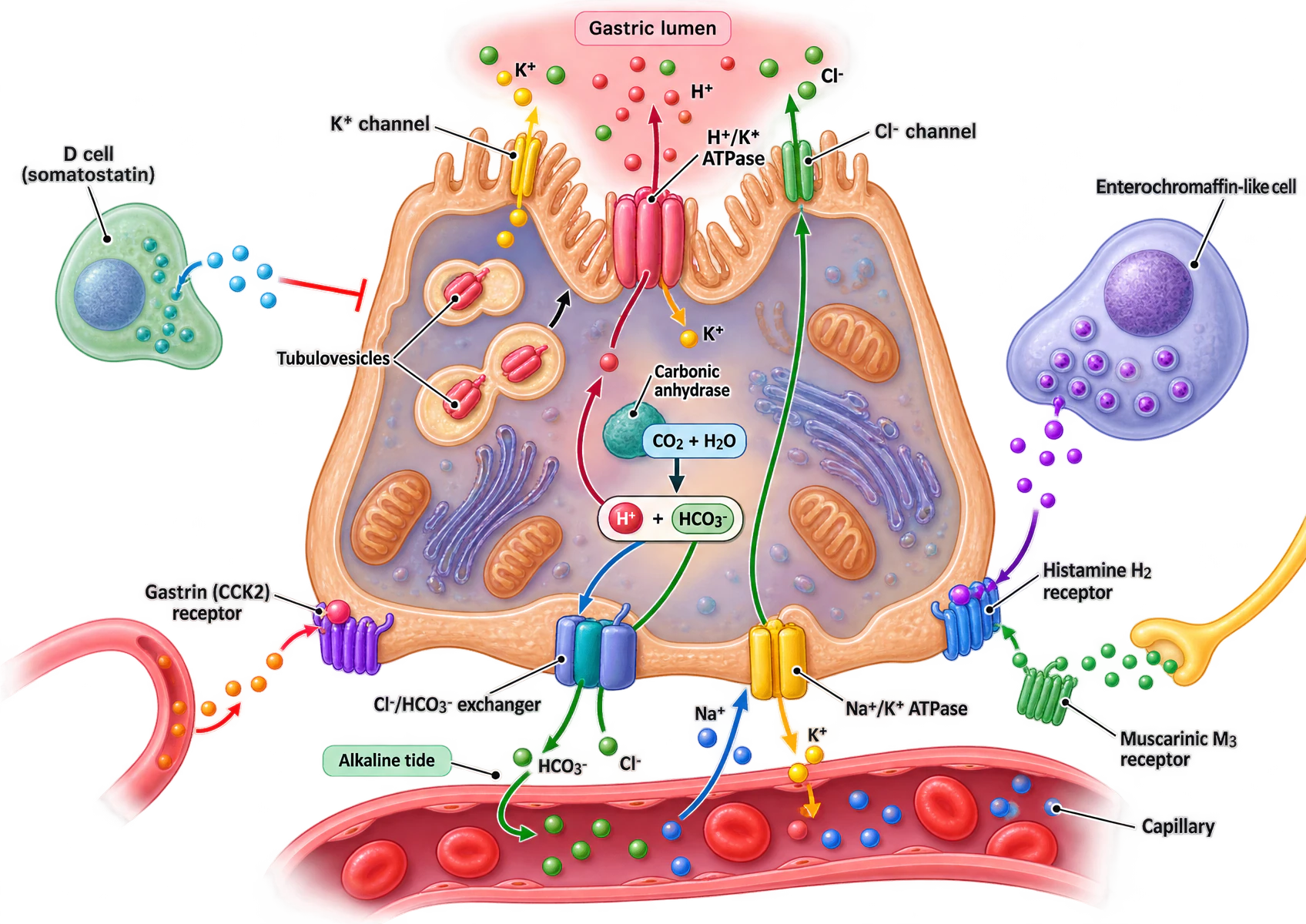
Two defects in the supplied artwork, neither affecting the mechanism. The secretory canaliculus is drawn but not labelled — it is the deep apical invagination lined with microvilli into which the pump secretes. And two labels render the potassium ion as “K*” rather than K+, in “K* channel” and “H+/K* ATPase”, although the hydrogen superscript in the same label is correct.
A single parietal cell is drawn in section, with the gastric lumen above and a capillary below. Its apical membrane is thrown into microvilli around a secretory canaliculus. In that apical membrane sit an H+/K+ ATPase, which exports hydrogen into the lumen in exchange for potassium, a potassium channel that returns potassium to the lumen to supply the pump, and a chloride channel through which chloride diffuses out, so that hydrochloric acid is the net luminal product. Tubulovesicles carrying more pump molecules are shown fusing with the apical membrane. In the cytoplasm, carbonic anhydrase converts carbon dioxide and water into hydrogen and bicarbonate ions. At the basolateral membrane, a chloride-bicarbonate exchanger sends bicarbonate into the capillary, labelled the alkaline tide, and takes up chloride; a sodium-potassium ATPase maintains the sodium and potassium gradients. Three stimulatory receptors sit in the basolateral membrane: a gastrin CCK2 receptor reached by gastrin arriving in the capillary, a histamine H2 receptor reached from an adjacent enterochromaffin-like cell, and a muscarinic M3 receptor reached from a vagal nerve terminal. A D cell at the left releases somatostatin, drawn as an inhibitory bar onto the cell.
The mechanism runs in four steps. Carbon dioxide diffuses into the cell from the blood. Carbonic anhydrase converts it with water to carbonic acid, which dissociates into H+ and HCO3−. At the apical membrane the H+/K+ ATPase — the proton pump — exports hydrogen into the secretory canaliculus in exchange for potassium, against a concentration gradient of roughly three million to one, which is why the cell is packed with mitochondria. At the basolateral membrane bicarbonate is exchanged for chloride; the bicarbonate enters the blood and the chloride crosses the cell and leaves through an apical chloride channel, so the net luminal product is hydrochloric acid and the net blood product is sodium bicarbonate.
| Receptor | Ligand and source | Second messenger | Importance |
|---|---|---|---|
| H₂ | Histamine, from the adjacent enterochromaffin-like cell (paracrine) | cAMP | The most important direct stimulus |
| M₃ | Acetylcholine, from vagal fibres through myenteric neurones | IP₃ and diacylglycerol, raising intracellular calcium | Directly stimulatory, and the mechanism of the cephalic phase |
| CCK₂ (gastrin) | Gastrin, arriving in the blood from antral G cells | IP₃ and diacylglycerol | The least important DIRECT stimulus, and the most important overall — because most of its effect is indirect, through histamine release |
| Somatostatin receptor | Somatostatin, from the local D cell (paracrine) | Inhibits adenylyl cyclase | The only inhibitory receptor. It is how luminal acid switches its own production off |
Secretion
The three phases of secretion
| Phase | Share | Stimulus | How it works |
|---|---|---|---|
| Cephalic phase | 30% | Thought, sight, smell and taste of food | Cortex and hypothalamus to the dorsal vagal nucleus, then vagus to the enteric plexuses. Acetylcholine directly on the parietal cell at M3, and gastrin-releasing peptide onto the G cell. Vagal activity also removes the somatostatin brake. |
| Gastric phase | 60% | Distension of the body and antrum, and peptides and amino acids in the lumen | Local enteric reflexes and vagovagal reflexes; gastrin into the portal blood. Gastrin, acting mostly indirectly by releasing histamine from enterochromaffin-like cells onto parietal H2 receptors. |
| Intestinal phase | 10% | Products of protein digestion in the duodenum, and amino acids in the blood | Duodenal endocrine cells and the enterogastric reflex. A small stimulatory contribution, quickly overtaken by inhibition: acid, fat and hyperosmolarity release secretin, gastric inhibitory peptide and somatostatin, which shut the parietal cell down. |
The negative feedback matters as much as the stimulation. Once acidic chyme reaches the duodenum, secretin is released and inhibits gastrin release from the G cells; gastric inhibitory peptide promotes somatostatin release, which inhibits both the G cell and the parietal cell directly; and the enterogastric reflex, carried by the enteric nervous system and by the extrinsic sympathetic and vagal supply, reduces both secretion and motility. That matching of delivery to the duodenum’s handling capacity is the single most important control in the section, and it is the same set of signals that will slow emptying in section 08.
Defence
Mucosal protection
The gastric mucosal barrier is not one thing. It has a pre-epithelial, an epithelial and a subepithelial component, and clinically useful damage requires more than one of them to fail.
| Level | Component | How it works | What defeats it |
|---|---|---|---|
| Pre-epithelial | The mucus-bicarbonate layer | An unstirred gel of mucus holds bicarbonate secreted by the surface cells against the epithelium, so a luminal pH of about 2 becomes a pH of about 7 at the cell surface across a few hundred micrometres | Anything that reduces mucus or bicarbonate secretion, chiefly loss of prostaglandin |
| Epithelial | Tight junctions and apical membranes | The surface cells are joined by tight junctions and their apical membranes are relatively impermeable to hydrogen ions | Bile salts and alcohol, which are lipid-soluble and cross the membrane directly |
| Epithelial | Rapid restitution | Surface cells are replaced every three to five days, and small defects are re-covered within minutes by neighbouring cells migrating across the gap | Anything that impairs cell turnover, including hypoperfusion |
| Subepithelial | Mucosal blood flow | Removes hydrogen ions that do cross, and delivers the bicarbonate and the oxygen the surface cells need to secrete it | Hypoperfusion — which is why stress ulceration is a disease of shock rather than of acid |
| Regulator | Prostaglandins, chiefly PGE₂ | Increase mucus and bicarbonate secretion and mucosal blood flow, and inhibit acid secretion. They regulate three of the four levels above at once | Non-steroidal anti-inflammatory drugs, by cyclo-oxygenase inhibition |
Secretion
Pepsinogen, intrinsic factor and mucus
| Secretion | Cell | Activated or acting how | Why it matters |
|---|---|---|---|
| Pepsinogen | Chief cell | Secreted as a proenzyme and cleaved autocatalytically to pepsin by the acid environment, optimally below pH 3. Irreversibly inactivated above pH 7 | Begins protein digestion, cleaving dietary protein to shorter polypeptides. Not essential: pancreatic proteases can complete the job alone |
| Intrinsic factor | Parietal cell | A glycoprotein that binds vitamin B₁₂ in the duodenum, where the pH has risen and where haptocorrin has been digested by trypsin. Absorbed as a complex by specific receptors in the terminal ileum | The only gastric secretion that is irreplaceable. Autoimmune destruction of parietal cells causes pernicious anaemia; so, eventually, does total gastrectomy |
| Mucus and bicarbonate | Mucous neck and surface cells | An adherent gel holding bicarbonate against the epithelium | Section 05. Also lubricates the wall against the mechanical damage of antral grinding |
| Gastric lipase | Chief cell | Acid-stable, unlike pancreatic lipase | Contributes modestly to fat digestion, and more in the neonate whose pancreatic lipase is immature |
About 2000 mL of gastric juice is produced a day. Its composition varies with the rate of secretion, because it is really two secretions mixed: a non-parietal component resembling plasma, and a parietal component of essentially pure hydrochloric acid at about 150 mmol/L. At high secretion rates the mixture approaches pure parietal secretion. That is why prolonged vomiting or nasogastric aspiration produces a hypochloraemic, hypokalaemic metabolic alkalosis with hypovolaemia: the fluid lost is rich in hydrogen, chloride and potassium, and its bicarbonate counterpart has already gone into the blood.
Motility
Motility and receptive relaxation
| Receptive relaxation | Accommodation | |
|---|---|---|
| Trigger | The bolus passing along the oesophagus, before it arrives | The presence of food already in the proximal stomach |
| Reflex arc | Vagovagal — the afferent and efferent limbs both run in the vagus | Local, through the enteric nervous system |
| Timing | Anticipatory | Adaptive, continuing as the meal is eaten |
| Transmitter | Nitric oxide and vasoactive intestinal peptide, from non-adrenergic non-cholinergic myenteric neurones | The same |
| Consequence of losing it | Early satiety and a rise in intragastric pressure on eating — seen after vagotomy and after fundoplication | The same |
The distal stomach does the opposite work. Its basal electrical rhythm is set by interstitial cells of Cajal on the greater curvature at about three cycles per minute, and that pacemaker drives the whole distal stomach at its own rate. Slow waves alone do not reach threshold: they determine when a contraction may occur, and vagal activity, gastrin and acetylcholine determine whether one does, by raising the plateau of the slow wave until it does reach threshold.
Motility
Gastric emptying
At two hours — the clear-fluid fasting interval — about 1.6% of a clear liquid remains, against about 50% of a solid meal. That difference, not a difference in risk of reflux, is why liquids may be taken so much closer to induction than solids.
| Factor | Effect | Mechanism |
|---|---|---|
| Gastric volume and distension | Accelerates | Distension provokes vagovagal excitatory reflexes and releases gastrin, raising antral pump activity |
| Consistency | Liquids leave far faster | The pylorus admits particles below 1 to 2 mm, so solids must be liquefied first |
| Composition | Carbohydrate fastest, then protein, then fat | Fat is slowest of all, acting through cholecystokinin released by fatty acids arriving in the duodenum |
| Duodenal distension | Slows | Reflex inhibition through the enteric nervous system |
| Duodenal acid | Slows | Hydrogen ions detected by duodenal chemoreceptors release secretin, which acts directly on gastric smooth muscle and reduces gastrin |
| Duodenal fat and protein products | Slows | Cholecystokinin raises pyloric tone, buying the small intestine time to digest the lipid already delivered |
| Duodenal hyperosmolarity | Slows | Duodenal osmoreceptors, through an enteric reflex. Isosmotic contents empty fastest |
| Sympathetic activity, pain, anxiety | Slows | Sympathetic inhibition of the enteric plexuses. This is why trauma and pain delay emptying regardless of when the patient last ate |
| Opioids | Slows markedly | Peripheral mu receptors in the myenteric plexus increase resting tone and abolish propulsive motility |
| Prokinetics | Accelerate | Metoclopramide and erythromycin, the latter a motilin agonist |
| Autonomic neuropathy, critical illness, raised intracranial pressure, hyperglycaemia | Slow | Loss of vagal drive, and of coordinated pacemaking |
Applied
Volume and pH of gastric contents
Fasting gastric juice has a pH of about 1 to 1.5 and the stomach secretes about 2000 mL a day. A fasted stomach is not an empty one: it always contains some residual volume of secretion, swallowed saliva and refluxed duodenal content, and that residual volume is what is available to be aspirated at induction.
The injury itself is Mendelson's syndrome: a chemical pneumonitis from the aspiration of acidic gastric contents. The mechanism is direct acid injury to the alveolar epithelium and capillary endothelium, producing an inflammatory exudate, surfactant loss, atelectasis and a large intrapulmonary shunt. Particulate material, even at a neutral pH, causes obstruction and a foreign-body reaction instead — which is why particulate antacids are avoided before induction and non-particulate sodium citrate is used: raising the pH is no help if the agent that raises it is itself damaging when aspirated.
Applied
Fasting and the full stomach
| Intake | Interval | Why |
|---|---|---|
| Clear fluids | 2 hours | Exponential emptying with no lag phase and a half-time of about 20 minutes, so about 1.6% remains at two hours. Carbohydrate drinks, and tea or coffee with up to a fifth of the volume as milk, count as clear fluids |
| Breast milk | 4 hours | Intermediate. Formula milk counts as solid food, because it curdles in the acid stomach |
| Solids | 6 hours | A lag phase then a linear fall with a half-time of about 2 hours, so a substantial fraction is still present at four |
A full stomach is a physiological state rather than a history. Treat the stomach as full, whatever the fasting interval, when emptying has been arrested or the volume is being added to: pain, trauma, opioids, an acute abdomen, bowel obstruction — in which the stomach is filling from below rather than emptying — labour, autonomic neuropathy, gastroparesis, raised intracranial pressure, and critical illness. In each of these the curves in section 08 simply do not apply, because the mechanism that generates them has been interrupted.
Consolidation
The lesson in one paragraph
The stomach stores 1.5 to 2 litres, mixes and grinds it to particles of 1 to 2 mm, and releases it slowly. Its proximal part is a reservoir producing tonic contractions; its distal part grinds with high-amplitude contractions paced by interstitial cells of Cajal at about three per minute, driving food back through a closing pylorus in retropulsion so that grinding is done by the food against itself. It accepts a meal without a rise in pressure by two reflexes with the same transmitters — receptive relaxation, which is vagovagal and anticipatory, and accommodation, which is local and adaptive, both mediated by nitric oxide and vasoactive intestinal peptide. Acid comes from the parietal cell, where carbonic anhydrase generates hydrogen and bicarbonate, the apical H+/K+ ATPase exports hydrogen against a gradient of about three million to one, and the basolateral chloride-bicarbonate exchanger returns bicarbonate to the blood as the alkaline tide. Three stimulatory receptors reach that pump — H2, M3 and gastrin at CCK2 — of which histamine is the most important direct stimulus and gastrin the most important overall, most of its effect being indirect through enterochromaffin-like cells; somatostatin from the D cell is the only inhibitory receptor and is how luminal acid switches off its own production. Secretion runs in three phases contributing about 30, 60 and 10 per cent of the acid: cephalic by vagal acetylcholine and gastrin-releasing peptide, gastric by distension and luminal peptides, intestinal briefly stimulatory and then the brake. The mucosa survives all of this behind a four-level barrier — the mucus-bicarbonate gel, tight junctions and impermeable apical membranes, rapid restitution, and mucosal blood flow — regulated at three levels by prostaglandins, which is why non-steroidal anti-inflammatory drugs and hypoperfusion are the two things that breach it and why stress ulceration is a disease of shock. Beside acid the stomach makes pepsinogen, activated autocatalytically below pH 3; gastric lipase; the mucus-bicarbonate layer; and intrinsic factor, the only irreplaceable gastric secretion, which binds vitamin B12 in the duodenum after haptocorrin is digested by trypsin and delivers it to receptors in the terminal ileum. About 2000 mL of juice is made daily at a fasting pH of 1 to 1.5, so prolonged vomiting or aspiration produces a hypochloraemic, hypokalaemic metabolic alkalosis with hypovolaemia. Emptying follows two different shapes: solids after a lag of 30 minutes then approximately linearly with a half-time of 2 hours, clear liquids exponentially from time zero with a half-time of 20 minutes — and it is the duodenum rather than the stomach that sets the rate, through distension, acid, fat and protein products, and hyperosmolarity. Those curves are what the 6, 4 and 2 hour fasting intervals encode, and they simply do not apply to the patient in pain, after trauma, on opioids, in labour, in obstruction or critically ill, whose stomach should be treated as full whatever the clock says.