What you should already have
About 75 minutes
Plus the time it takes to redraw this lesson’s figures from memory, which is the fastest way to find out what you have not understood.
Where this shows up
Almost everything you do to an anaesthetised patient reduces hepatic blood flow, and the liver has one compensating mechanism which several of those things also blunt. This lesson is also where the zonation comes from — and zonation is why paracetamol, halothane and shock all injure the same part of the lobule.
Learning outcomes
By the end of this lesson you should be able to:
- Contrast the lobule with the acinus, and place zones 1, 2 and 3 correctly relative to the portal triad and the central vein.
- State the oxygen tension at each end of the sinusoid and derive the metabolic zonation and the pattern of centrilobular injury from it.
- Describe the sinusoid and the space of Disse, including the fenestrations, the stellate cell and the Kupffer cell.
- Quantify the dual blood supply: total flow, the share of cardiac output, the flow and oxygen saturation of each vessel, and the share of oxygen each delivers.
- Describe the hepatic arterial buffer response, its mediator, the size of the compensation and why it works in one direction only.
- State the intrinsic controls of hepatic blood flow, including the pressure below which hepatic arterial autoregulation fails.
- Give the extrinsic controls: sympathetic innervation, the capacitance response, and the vasoactive substances that act on each vessel.
- State the effect on hepatic blood flow of spontaneous ventilation, positive-pressure ventilation and PEEP, hypocapnia and hypercapnia, regional and general anaesthesia, and surgery.
- Define hepatic extraction ratio and hepatic clearance, and explain when clearance measures liver blood flow and when it measures enzyme activity.
- Name the methods of measuring hepatic blood flow and the assumption each makes.
Together these settle one syllabus objective: Hepatic structure and zonation, the dual blood supply, the control of hepatic blood flow, and hepatic clearance. Tick it on the Physiology objective list once you can do all of the above without notes.
Orientation
Rapid review
- 1500 mL/min, about 25% of cardiac output. Split roughly a quarter to a third arterial and the remainder portal.
- Each vessel delivers about half the oxygen. The artery carries the minority of the blood at 98 to 100% saturation; the portal vein carries the majority at 85 fasting, about 70 fed%. That asymmetry is the single most examinable fact in the section.
- The acinus is not the lobule. The lobule is the anatomical unit with a central vein in the middle; the acinus is the functional unit with the portal triads in the middle and a venule at each end. Zones run 3-2-1-2-3 across it.
- Zone 1 is periportal and best oxygenated; zone 3 is perivenous and worst. 60–65 mmHg falling to 30–35 mmHg. Zone 3 does the drug metabolism and dies first.
- The hepatic arterial buffer response is semi-reciprocal. A fall in portal flow raises arterial flow by 22 to 100%, mediated by adenosine washout. A fall in arterial flow is not compensated at all.
- The buffer is partial. Total hepatic flow still falls. The artery defends it; it does not restore it.
- Surgery beats anaesthesia. Retraction and packing reduce hepatic blood flow more than any anaesthetic factor.
Structure
The lobule, the acinus and zonation
The hepatic lobule, and the sinusoid in detail
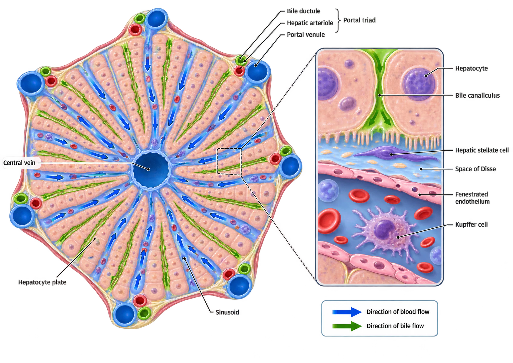
Three defects in the supplied artwork. One sinusoid arrow in the upper left of the lobule points outwards; blood in every sinusoid flows inwards, from the portal triads at the corners to the central vein. The canaliculi in the lobule panel carry no arrowheads, so the key’s green bile arrow has nothing there to refer to. And in the inset the bile arrowheads run down the canaliculus towards the sinusoid; bile drains the other way, towards the portal triad, and the canaliculus is sealed from the space of Disse by tight junctions. Bile never enters the sinusoid in health.
A hexagonal hepatic lobule seen in section. A central vein sits at the middle; six portal triads sit at the corners, each drawn as three vessels — a blue portal venule, a red hepatic arteriole and a green bile ductule. Plates of hepatocytes radiate from the centre to the periphery, with blood-filled sinusoids between them and blue arrows in the sinusoids indicating flow from the corners inwards to the central vein. Green channels running along the hepatocyte plates represent the bile canaliculi. To the right, an inset magnifies a single sinusoid: two hepatocytes with a bile canaliculus in the cleft between them, a perisinusoidal space of Disse containing a hepatic stellate cell, a fenestrated endothelial lining, and a Kupffer cell within the sinusoidal lumen among red cells. A key gives a blue arrow for blood flow and a green arrow for bile flow.
| Classical lobule | Acinus | |
|---|---|---|
| Kind of unit | Anatomical, or structural | Functional |
| Shape | Hexagonal | Elliptical or diamond-shaped |
| At the centre | The central vein | Two portal triads, at the midpoints of the long sides |
| At the periphery | Six portal triads, one at each corner | A terminal hepatic venule at each end |
| Blood flows | From the corners inwards to the centre | From the centre outwards to both ends |
| Why it exists | It is what the tissue looks like down a microscope | It is organised around the oxygen gradient, so it predicts function and injury |
| Zone | Position | Oxygen tension | Predominant processes | Vulnerability |
|---|---|---|---|---|
| Zone 1 — Periportal | Closest to the portal triads, at the midpoints of the acinus | 60 to 65 mmHg | Aerobic metabolism and oxidative phosphorylation; Gluconeogenesis, and glycogen synthesis from lactate and amino acids; Urea synthesis from ammonia; Beta-oxidation of fatty acids; Bile acid synthesis; Sulfation | First to meet an ingested toxin arriving in portal blood, and the last to become hypoxic. |
| Zone 2 — Intermediate | Between the two | Intermediate | Processes characteristic of both neighbouring zones | No distinctive pattern of its own. |
| Zone 3 — Perivenous, pericentral or centrilobular | Closest to the terminal hepatic venules at the ends of the acinus | 30 to 35 mmHg | Glycolysis, and glycogen synthesis from glucose; Lipogenesis and ketogenesis; Cholesterol synthesis; Glutamine synthesis, the high-affinity ammonia scavenger; Glucuronidation; Cytochrome P450 drug metabolism | Lowest oxygen tension, so the first to die in hypoperfusion, and the site of centrilobular necrosis in shock, in paracetamol overdose and in halothane hepatitis. |
Structure
The sinusoid and the space of Disse
The sinusoid is a specialised capillary in which mixed portal and arterial blood flows past the hepatocytes. Its endothelium is fenestrated, with pores of roughly 50 to 150 nm and no basement membrane, so plasma, plasma proteins, lipoproteins and drug molecules pass freely into the space of Disse between the endothelium and the hepatocyte, while blood cells are retained. That is why the hepatocyte is exposed to essentially the whole plasma composition rather than to a filtrate, and it is the structural reason hepatic extraction can be as efficient as it is.
| Cell | Share | Where | What it does |
|---|---|---|---|
| Hepatocyte | 75 to 80% of cellular volume | In plates, with a basolateral surface facing the space of Disse and an apical surface forming the bile canaliculus | Everything in lesson 6 |
| Sinusoidal endothelial cell | — | Lining the sinusoid | Fenestrated, with no basement membrane, so solutes reach the space of Disse freely; larger molecules cross by transcytosis |
| Kupffer cell | 20 to 30% of the non-parenchymal cells, and 80 to 90% of all the body's tissue macrophages | Within the sinusoidal lumen | Phagocytose bacteria and endotoxin arriving in portal blood, before they reach the systemic circulation |
| Hepatic stellate cell | 8 to 10% of resident liver cells | In the space of Disse | Quiescent in health, storing vitamin A. On injury they transform into myofibroblasts and lay down collagen — the central event in hepatic fibrosis |
Blood supply
The dual blood supply
The dual supply, and the buffer response in the portal tract
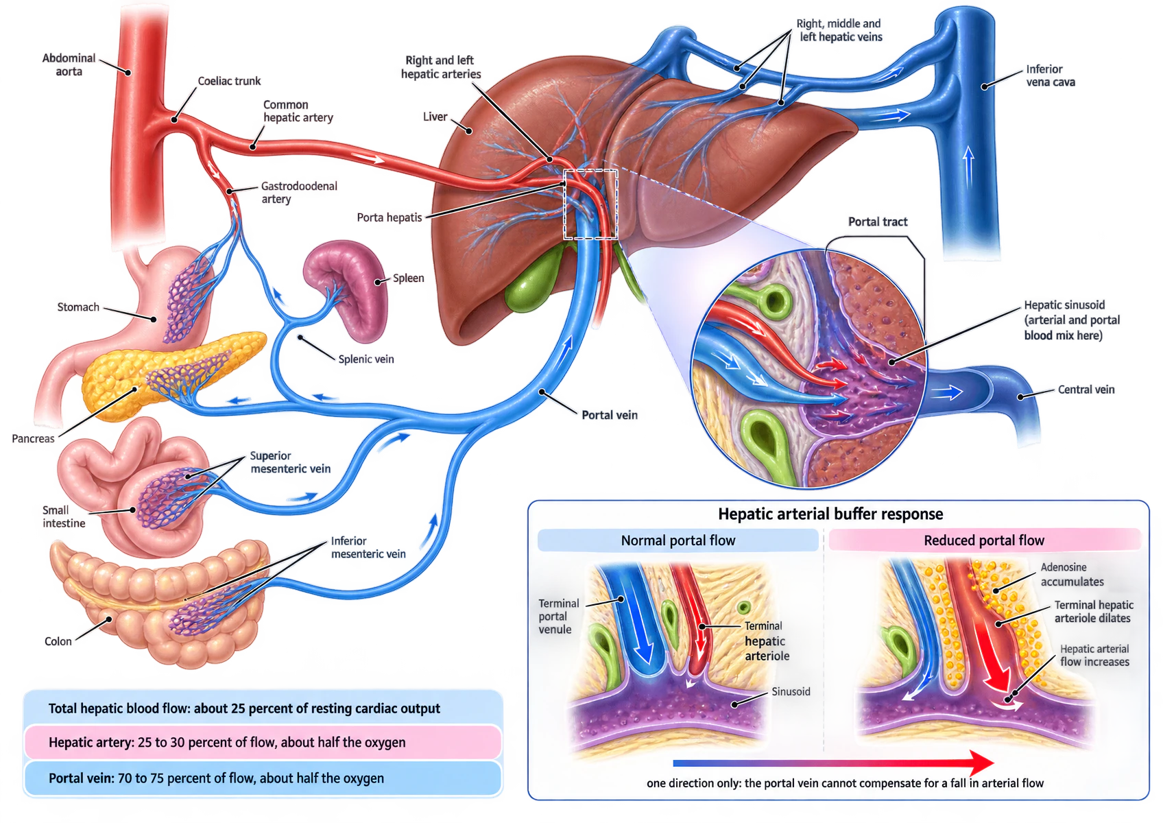
One defect in the supplied artwork: the vessel labelled “Gastrodoodenal artery” is the gastroduodenal artery. The spelling is an error in the export and cannot be corrected in the file. Everything else in the figure, including the buffer-response panel, is accurate.
A schematic of the hepatic circulation. From the abdominal aorta the coeliac trunk gives the common hepatic artery, which gives off the gastroduodenal artery and then enters the porta hepatis dividing into right and left hepatic arteries. Below, the stomach, pancreas, spleen, small intestine and colon drain through the splenic, superior mesenteric and inferior mesenteric veins into a single portal vein, which also enters the porta hepatis. Right, middle and left hepatic veins leave the liver for the inferior vena cava. A circular inset magnifies a portal tract and sinusoid: a red terminal hepatic arteriole and a blue terminal portal venule both open into a purple sinusoid where the two bloods mix, and the sinusoid drains to a central vein. A lower panel compares normal portal flow with reduced portal flow: in the second, yellow dots labelled adenosine accumulate around the terminal hepatic arteriole, which dilates, and hepatic arterial flow increases. A bar beneath states that the relationship works in one direction only, and that the portal vein cannot compensate for a fall in arterial flow. Three coloured panels give total hepatic blood flow as about 25 per cent of resting cardiac output, the hepatic artery as 25 to 30 per cent of flow and about half the oxygen, and the portal vein as 70 to 75 per cent of flow and about half the oxygen.
| Hepatic artery | Portal vein | |
|---|---|---|
| Flow | 500 mL/min, 25 to 33% of the total | 1000 mL/min, 67 to 75% of the total |
| Oxygen delivered | 40 to 50% | 50 to 60% |
| Oxygen saturation | 98 to 100% | 85 fasting, about 70 fed% |
| Pressure | Systemic; about 35 mmHg in the hepatic arteriole | 5 to 10 mmHg |
| Origin | Coeliac trunk in about 80%; from the superior mesenteric artery in the remainder | Superior mesenteric, splenic and inferior mesenteric veins |
| Autoregulates | Yes, to a degree | No — it has almost no smooth muscle, and flow is proportional to the pressure gradient |
| Can compensate for the other | Yes — the buffer response | No |
Control
The hepatic arterial buffer response
The mechanism is adenosine washout, and it is elegant because it needs no sensor at all. Adenosine is produced continuously at a constant rate into the small fluid space of the portal tract, which contains the terminal portal venule and the terminal hepatic arteriole together. Portal blood flowing past washes it away. When portal flow falls, less adenosine is removed, so its local concentration rises; adenosine is a potent vasodilator, and the hepatic arteriole beside it dilates. Nothing measures anything: the concentration of the mediator is itself the signal, because it is set by the flow that is being sensed.
Control
Intrinsic control
| Mechanism | Acts on | How | Limit |
|---|---|---|---|
| The hepatic arterial buffer response | The hepatic artery | Adenosine washout: reduced portal flow leaves adenosine to accumulate, dilating the arteriole | An arterial rise of 22 to 100%, and it does not operate in reverse |
| Autoregulation | The hepatic artery only | Arteriolar resistance falls as perfusion pressure falls, holding flow relatively constant. A myogenic mechanism, weaker than in the brain or the kidney and best demonstrated in the fed state | Fails below a mean arterial pressure of about 60 mmHg — one source frames the same limit as a systolic pressure of 80 mmHg — below which flow becomes pressure-passive |
| Pressure-flow, with no regulation | The portal vein | The portal vein has very little smooth muscle. Flow is simply proportional to the pressure gradient across the liver, and is set by the mesenteric arterioles upstream | There is no limit because there is no regulation. This is the whole reason the buffer response is needed |
| The hepatic venous pressure effect | The hepatic artery | A rise in hepatic venous pressure raises hepatic arterial resistance, probably myogenically | Which is why congestive cardiac failure, PEEP and a raised intra-abdominal pressure all reduce arterial flow as well as impeding outflow |
Control
Extrinsic control
| Influence | Hepatic artery | Portal vein and capacitance vessels |
|---|---|---|
| Receptors present | Alpha and beta adrenoceptors, and dopamine receptors | Alpha adrenoceptors and dopamine receptors only |
| Sympathetic stimulation | Vasoconstriction | Venoconstriction, expelling stored blood into the circulation — the capacitance function of lesson 1 |
| Adrenaline | Initial alpha-mediated constriction, then beta-mediated dilatation | Constriction |
| Angiotensin II | Constricts | Constricts |
| Vasopressin | Constricts | Constricts and reduces portal flow — which is why it and its analogues are used in variceal bleeding |
| Glucagon | Dilates, raising hepatic blood flow | — |
| Secretin and vasoactive intestinal peptide | Dilate | Minimal effect |
| Feeding | — | Raises mesenteric flow and therefore portal flow substantially, which is the largest physiological increase in hepatic blood flow there is |
Two systemic states deserve separate mention. Vigorous exercise constricts the splanchnic bed and reduces hepatic blood flow. Graded hypoxia initially reduces hepatic arterial flow, which then returns to baseline within about twenty minutes, with minimal effect on portal flow; hyperoxia has little effect on either. Neither is a regulatory mechanism so much as a consequence of what the rest of the circulation is doing, which is the general pattern for extrinsic control here.
Applied
Ventilation, anaesthesia and surgery
| State | Effect | Mechanism | The buffer response |
|---|---|---|---|
| Spontaneous ventilation | Preserved | Mean intrathoracic pressure stays negative, so the pressure gradient from the hepatic veins to the right atrium is maintained and venous return is not impeded. Within a single breath the sources disagree about the direction of the phasic change; the mean is what matters. | Intact |
| Positive-pressure ventilation | Reduced | Raised mean intrathoracic pressure reduces venous return and cardiac output, and portal flow falls with it. Kam attributes the fall principally to the fall in cardiac output rather than to a direct hepatic effect. | Intact, and partially defends total flow |
| PEEP | Reduced | Adds to mean intrathoracic pressure through the whole respiratory cycle, so it does the same thing as positive-pressure ventilation, continuously. Also raises hepatic venous pressure, which itself raises hepatic arterial resistance. | Blunted, because a raised hepatic venous pressure constricts the artery |
| Hypocapnia | Reduced | Reduces hepatic blood flow by about 30%, mainly by raising resistance in the portal system. A consequence of over-ventilating, and therefore avoidable. | Intact |
| Hypercapnia | Increased | Increases hepatic blood flow, through an increase in portal venous flow. | Not called upon |
| Regional anaesthesia | Reduced | Epidural blockade reduces total hepatic blood flow, largely through reduced portal venous flow and reduced mean arterial pressure. Four of the five studies reviewed found a reduction, measured by indocyanine green plasma disappearance rate and transgastric hepatic vein Doppler, and restoring the pressure with ephedrine or noradrenaline reduced flow further rather than restoring it. The fifth found thoracic blockade increased flow while lumbar blockade reduced it, so the site of the block may matter and the clinical significance is not settled. | Intact |
| Volatile anaesthesia | Reduced | Reduces mean arterial pressure and cardiac output dose-dependently, and portal flow falls with them. Isoflurane, sevoflurane and desflurane preserve the buffer response, so total flow and hepatic oxygenation are largely maintained; halothane does not. | Preserved with isoflurane, sevoflurane and desflurane. Abolished by halothane. |
| Intravenous anaesthesia | Reduced | Thiopentone, etomidate and propofol reduce hepatic blood flow dose-dependently through reduced cardiac output, and Kam adds obtundation of the buffer mechanism. Miller records the opposite finding for propofol in studies using microspheres and Doppler, and attributes the disagreement to propofol interfering with the indocyanine green assay itself. | Reported both ways; see the lesson |
| Surgery: retraction and packing | Markedly reduced | Direct mechanical interruption of inflow and outflow. Chambers states plainly that intraoperative retraction and packing reduce hepatic blood flow significantly more than any anaesthetic factor. | Cannot compensate for a mechanical obstruction |
| Surgery: pneumoperitoneum | Reduced | Raised intra-abdominal pressure compresses the portal vein and the hepatic veins, and raises hepatic venous pressure, which itself raises hepatic arterial resistance. | Blunted |
| Surgery: the stress response and haemorrhage | Reduced | Sympathetic activity constricts the splanchnic bed, so portal flow falls before arterial pressure does. In acute haemorrhage portal flow falls more than arterial flow, and hepatic oxygen supply is defended by increased extraction rather than by increased flow. | Intact, and this is when it matters most |
Applied
Hepatic clearance and the extraction ratio
where Q is hepatic blood flow (about 1500 mL/min) and ER is the hepatic extraction ratio, the fraction of drug removed in a single pass. A high extraction ratio is conventionally above 0.7 and a low one below 0.3, with an intermediate band between.
The circulatory reading is the one this lesson adds. Everything in section 08 moves Q. For a high-extraction drug — propofol, lignocaine, propranolol, morphine — that means every one of those states changes its clearance directly, which is why a propofol infusion behaves differently in a shocked patient and why lignocaine toxicity is a risk in cardiac failure. For a low-extraction drug — warfarin, phenytoin, theophylline, diazepam — none of it matters much, and what does matter is enzyme induction or inhibition and protein binding.
Applied
Measuring hepatic blood flow
| Method | Principle | The assumption it makes | Limitation |
|---|---|---|---|
| Clearance techniques | The Fick principle applied to a substance the liver alone extracts avidly. Indocyanine green is the usual marker, with an extraction ratio of about 0.74; propranolol, lignocaine and radiolabelled colloids have also been used. Flow = clearance ÷ extraction ratio | That hepatocyte function is normal, and that the marker is extracted exclusively by the liver | Precisely the assumption that fails in liver disease, which is when you most want the measurement. A fall in indocyanine green clearance may mean reduced flow, reduced hepatocyte function, or both, and cannot distinguish them |
| Indicator dilution | A known quantity of a radiolabelled indicator that resists hepatic clearance is injected, and its concentration measured continuously from a hepatic vein. Flow is calculated from the dilution curve | That hepatic perfusion is uniform | Works in the presence of liver dysfunction, unlike clearance methods, but is invalidated by intrahepatic or extrahepatic shunting. Invasive, and a research tool |
| Direct measurement | Electromagnetic or ultrasonic flow probes placed around the hepatic artery or portal vein at laparotomy; implantable Doppler probes after transplantation | That placing the probe has not changed what it is measuring | It usually has: the surgery and anaesthesia required to implant it themselves reduce hepatic blood flow. Clinically used mainly to detect hepatic artery or portal vein thrombosis after transplantation |
A fourth family measures hepatic function rather than flow and is easily confused with these — the indocyanine green plasma disappearance rate, caffeine clearance, the aminopyrine breath test, monoethylglycinexylidide from lignocaine. The confusion is not accidental: because indocyanine green is highly extracted, its clearance depends on flow and on hepatocyte function together, which is exactly the extraction-ratio arithmetic of section 09 read from the other end.
Consolidation
The lesson in one paragraph
The liver receives about 1500 mL/min, roughly 25% of the cardiac output, down two inflows that differ in every respect. The hepatic artery brings 25 to 33% of the blood at 98 to 100% saturation and systemic pressure, and 40 to 50% of the oxygen; the portal vein brings the remainder at 85 fasting, about 70 fed% saturation and 5 to 10 mmHg, and about the same share of the oxygen — a minority of the blood carrying half the oxygen. Both drain into sinusoids at only 2 mmHg, lined by fenestrated endothelium with no basement membrane, so plasma reaches the hepatocyte across the space of Disse, where the stellate cells sit that produce fibrosis and capillarise the sinusoid when they are activated. Structurally the tissue is a lobule, hexagonal with a central vein at its centre; functionally it is an acinus, with the portal triads at the centre and a terminal venule at each end, so the zones read 3-2-1-2-3 and zone 1 is periportal and best oxygenated at 60–65 mmHg while zone 3 is perivenous at 30–35. Zone 1 does gluconeogenesis, urea synthesis and beta-oxidation; zone 3 does glycolysis, lipogenesis, glutamine synthesis and cytochrome P450 metabolism, and dies first in shock, in paracetamol overdose and in halothane hepatitis. Flow is controlled intrinsically by three things: weak hepatic arterial autoregulation that fails below a mean pressure of about 60 mmHg; no portal autoregulation at all, portal flow being simply proportional to the pressure gradient and set upstream; and the hepatic arterial buffer response, in which a fall in portal flow lets constantly produced adenosine accumulate in the portal tract and dilate the adjacent arteriole, raising arterial flow by 22 to 100%. It is semi-reciprocal and partial: the vein cannot compensate for the artery, and total flow still falls. Extrinsically the artery carries alpha, beta and dopamine receptors and the portal vein only alpha and dopamine, sympathetic activity constricting both and expelling the capacitance volume of lesson 1. Almost everything done to an anaesthetised patient reduces hepatic blood flow — positive-pressure ventilation and PEEP by raising intrathoracic pressure, hypocapnia by about 30% through portal resistance, neuraxial and general anaesthesia by reducing pressure and cardiac output — with surgical retraction and packing the largest single reduction of all, and halothane the one volatile that abolishes the buffer rather than preserving it. Spontaneous ventilation preserves flow relative to positive-pressure ventilation on the mean, though the sources contradict each other on the direction within a single breath. Finally, because Cl = Q × ER, all of that moves the clearance of a high-extraction drug directly and leaves a low-extraction drug almost untouched.