What you should already have
About 70 minutes
Plus the time it takes to redraw this lesson’s figures from memory, which is the fastest way to find out what you have not understood.
Where this shows up
This is the physiology behind every rapid sequence induction you will ever do. It is also the physiology that explains why a patient who has fasted correctly can still aspirate, why the drug you reach for at induction makes the problem worse, and why treating reflux successfully tells you nothing at all about whether the barrier is intact.
Learning outcomes
By the end of this lesson you should be able to:
- Describe the three phases of swallowing, naming which is voluntary, which nerves carry the afferent and efferent limbs, and where the reflex is coordinated.
- List the mechanisms that protect the airway during the pharyngeal phase, and give the duration of deglutition apnoea.
- Contrast the upper and lower oesophageal sphincters on structure, innervation, resting pressure and whether each is anatomical or physiological.
- Define barrier pressure, state its normal value, and explain why competence can be lost with sphincter tone unchanged.
- Classify the factors that alter lower oesophageal sphincter tone as mechanical, neural, hormonal, luminal and state-dependent, and give the direction of each.
- Explain transient lower oesophageal sphincter relaxations as normal vagal physiology, and say why they matter more than resting tone in obesity.
- Describe the gastrointestinal changes of pregnancy that reduce barrier pressure, and separate the hormonal from the mechanical contribution.
- State what anaesthetic agents, neuromuscular blockers and cricoid pressure do to the barrier, including the two drugs that raise tone.
Together these settle one syllabus objective: Swallowing, the oesophageal sphincters, barrier pressure and the physiological factors preventing reflux. Tick it on the Physiology objective list once you can do all of the above without notes.
Orientation
Rapid review
- Three phases, one voluntary. Oral (voluntary), pharyngeal (involuntary), oesophageal (involuntary). The reflex is coordinated in the swallowing centre of the medulla; afferents arrive by the glossopharyngeal and vagus nerves and efferents leave by the nucleus ambiguus.
- The airway is closed, not merely covered. The vocal cords adduct, the aryepiglottic folds close, the larynx is pulled up and forward under the tongue base, and only then does the epiglottis tilt over the inlet. Breathing stops for 1 to 2 seconds.
- Two sphincters, and only one is anatomical. The upper is cricopharyngeus, a named skeletal muscle. The lower has no discrete muscle to dissect and is identified by a high-pressure zone on manometry, which is what “physiological sphincter” means.
- Two muscular components, two different nerves. Intrinsic oesophageal smooth muscle on the vagus, and the crural diaphragm on the phrenic. An answer naming one has described half the sphincter.
- Barrier pressure = sphincter pressure − intragastric pressure. About 15 to 25 mmHg. Competence is the difference, so it falls either because the sphincter weakens or because the stomach pushes harder.
- Transient relaxations are normal. Vagally mediated, unrelated to swallowing, triggered by fundal distension, and the route by which most physiological reflux occurs. They are the dominant mechanism of reflux in obesity, not a low resting tone.
Deglutition
The oral and pharyngeal phases
A swallow, panel by panel
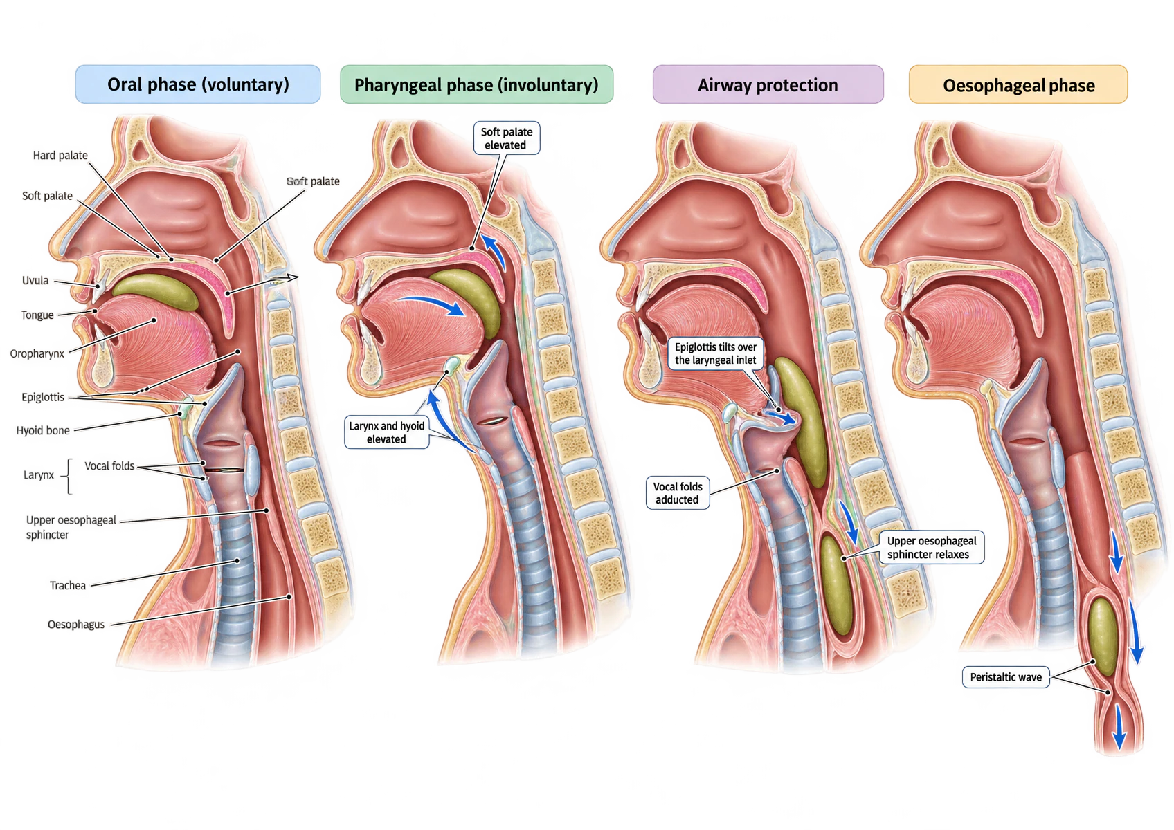
Three defects in the supplied artwork, none of which changes the physiology. Panel 1 carries the label “Soft palate” twice. In panel 3, the “Upper oesophageal sphincter relaxes” leader ends on the bolus rather than on the sphincter itself. And the figure is named for three phases but draws four panels: airway protection is an expansion of the pharyngeal phase, not a fourth phase of its own.
Four mid-sagittal sections of the head and neck. Panel one, the oral phase, is fully labelled: hard palate, soft palate, uvula, tongue, oropharynx, epiglottis, hyoid bone, larynx with its vocal folds, upper oesophageal sphincter, trachea and oesophagus, with the bolus held against the hard palate by the tongue. Panel two, the pharyngeal phase, shows the soft palate elevated to close the nasopharynx and the hyoid and larynx pulled upwards and forwards, with arrows marking both movements. Panel three shows airway protection: the epiglottis tilted down over the laryngeal inlet, the vocal folds adducted, and the upper oesophageal sphincter relaxing to admit the bolus. Panel four, the oesophageal phase, shows the bolus descending the oesophagus with arrows marking the peristaltic wave behind it.
| Phase | Control | What happens | Nerves |
|---|---|---|---|
| Oral | Voluntary | The tongue forms the bolus and drives it up against the hard palate and back into the oropharynx. Receptors in the posterior pharyngeal wall and soft palate then trigger the reflex, and from that moment the sequence cannot be interrupted. | Afferent: glossopharyngeal. Efferent: hypoglossal to the tongue, trigeminal to the muscles of mastication |
| Pharyngeal | Involuntary | The nasopharynx is closed by the soft palate, the larynx is closed and elevated, the pharyngeal constrictors contract in sequence, and cricopharyngeus relaxes and then tightens behind the bolus. Respiration is inhibited throughout. | Afferent: glossopharyngeal and vagus to the nucleus tractus solitarius. Efferent: nucleus ambiguus by vagus and glossopharyngeal |
| Oesophageal | Involuntary | The upper sphincter closes behind the bolus and the lower sphincter relaxes ahead of it. A primary peristaltic wave carries the bolus down; a secondary wave clears anything left behind. | Vagus for the primary wave; the enteric nervous system alone for the secondary wave |
Deglutition
How the airway is protected
The commonest wrong answer here is “the epiglottis covers the larynx”, as though that were the mechanism rather than the last step of it. Patients whose epiglottis has been removed can still swallow safely. What actually protects the airway is a sequence, and it works from the inside out.
- The vocal cords adduct, closing the glottis. Lateral cricoarytenoid, and the oblique and transverse arytenoids, all supplied by the recurrent laryngeal nerve.
- The aryepiglottic folds adduct, closing the laryngeal inlet above the cords. A second seal, above the first.
- The larynx and hyoid are pulled upwards and forwards, by the digastric and stylohyoid. This tucks the laryngeal inlet under the base of the tongue and out of the bolus’s path, and it simultaneously opens the upper oesophageal sphincter by traction.
- The epiglottis tilts down over the inlet, deflecting the bolus laterally into the piriform fossae. This is a diversion, not a lid.
- Respiration is inhibited for 1 to 2 seconds. Deglutition apnoea is brief enough to go unnoticed, and it means that no inspiratory effort can draw the bolus towards the cords at the moment they are most exposed.
Deglutition
The oesophageal phase
| Primary peristalsis | Secondary peristalsis | |
|---|---|---|
| Trigger | The swallow itself | Distension of the oesophageal wall by a bolus left behind |
| Generated by | The medullary swallowing centre, through the vagus | The enteric nervous system alone |
| Runs | From the top of the oesophagus to the lower sphincter, whether or not there is a bolus in front of it | From wherever the residual bolus is |
| Purpose | Delivery | Clearance, including of refluxed material |
| Survives vagotomy | No, in the striated upper third | Yes |
Two velocities are worth carrying because they explain why the pharyngeal phase feels instantaneous and the oesophageal phase does not. The bolus crosses the pharynx at about 30 cm/s and the oesophagus at about 3 cm/s. Gravity moves fluids faster than this in the upright position, which is why a drink reaches the stomach before the peristaltic wave does, and why swallowing works upside down but more slowly.
The sphincters
The upper oesophageal sphincter
The upper oesophageal sphincter is cricopharyngeus, the lowest and most horizontal part of the inferior constrictor, reinforced by the inferior constrictor proper and the circular muscle of the upper oesophagus. It lies at the level of the cricoid cartilage. It is skeletal muscle, so it is an anatomical sphincter in a way the lower one is not: you can find it, name it and dissect it.
Its job is bidirectional. It stops air entering the oesophagus during inspiration, and it stops oesophageal contents entering the pharynx. It relaxes during the pharyngeal phase — partly by active inhibition, partly by the traction of laryngeal elevation — and then contracts behind the bolus. Because it is skeletal muscle it has tone only while the patient is awake: it relaxes in sleep and under anaesthesia, which is why regurgitated material that reaches the oesophagus of an anaesthetised patient meets no upper barrier at all.
The sphincters
The lower oesophageal sphincter
The gastro-oesophageal junction, and the two pressures that meet there
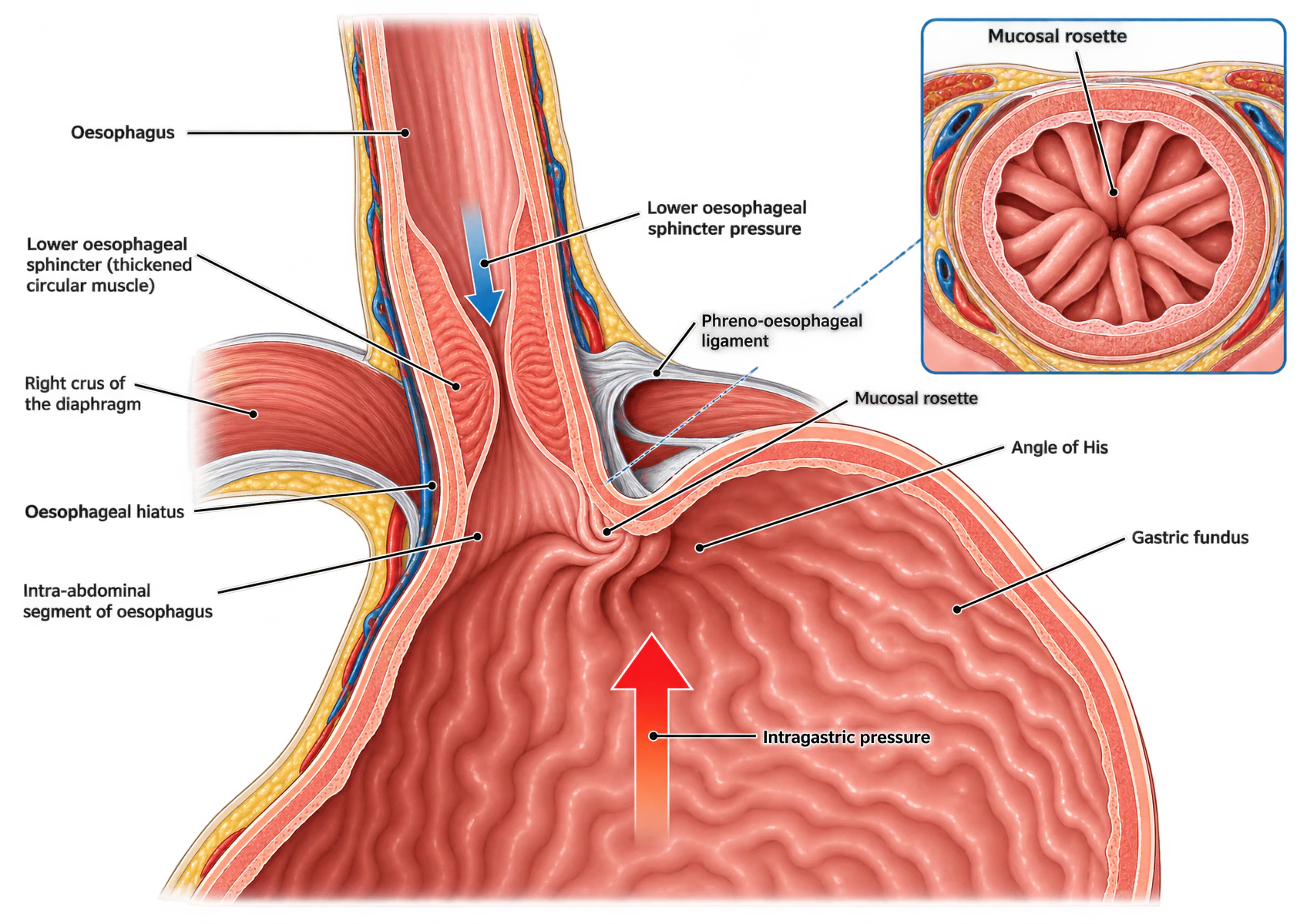
A coronal section through the gastro-oesophageal junction. The oesophagus descends from the top, passing through the oesophageal hiatus in the diaphragm, where the right crus is shown encircling it as a sling. The phreno-oesophageal ligament is drawn anchoring the oesophagus to the diaphragm. Below the hiatus, the intra-abdominal segment of oesophagus enters the stomach at the angle of His, an acute angle between the oesophagus and the gastric fundus, with the mucosal rosette at the junction itself. A blue arrow within the lumen points downwards, labelled lower oesophageal sphincter pressure; a red arrow rising from the gastric lumen points upwards, labelled intragastric pressure. An inset panel at upper right shows the mucosal rosette in cross-section as radiating folds occluding the lumen.
| Component | What it is | Nerve | What it contributes |
|---|---|---|---|
| Intrinsic sphincter | Tonically contracted circular smooth muscle of the distal 2 to 4 cm | Vagus | The resting tone, which is largely myogenic and increases when the muscle is stretched |
| Crural diaphragm | The right crus, slung around the oesophagus at the hiatus | Phrenic | A phasic external pinch that tightens on inspiration and on any expulsive effort — a feed-forward mechanism, since the act that raises the challenge also raises the defence |
| Angle of His | The acute angle at which the oesophagus enters the fundus | — | A flap valve, reinforced as the fundus distends |
| Intra-abdominal segment | The short length of oesophagus below the hiatus | — | Exposed to intra-abdominal pressure, so it is compressed by the same pressure that would otherwise force contents up it |
| Phreno-oesophageal ligament | The fascial attachment of oesophagus to diaphragm | — | Holds the intrinsic sphincter at the level of the crus, so the two components act together |
| Mucosal rosette | Redundant mucosal folds at the junction | — | Occludes the last millimetre of lumen |
The five-component list matters more than any single one of them, because it is what makes a hiatus hernia intelligible. A hiatus hernia removes three of the five at once: the intrinsic sphincter is displaced above the hiatus so it no longer acts at the same level as the crus, the intra-abdominal segment is lost, and the angle of His is straightened. The muscle itself may be entirely normal.
A swallow-related relaxation is a different event. The sphincter relaxes 1 to 2 seconds after the swallow begins, stays relaxed for 8 to 9 seconds, and then contracts 1 to 15 mmHg above its resting tone for 10 to 15 seconds before settling. Both relaxations are mediated by the same transmitters: nitric oxide and vasoactive intestinal peptide, released from the inhibitory non-adrenergic non-cholinergic neurones of the myenteric plexus.
Control
What sets and changes sphincter tone
Resting tone is largely myogenic: the muscle maintains a contractile state independent of neural input, and increases it when stretched. Everything below modulates that baseline. The five groups are the structure of the answer, and giving both directions within each group matters more than the length of any one list.
Mechanical
| Factor | Direction | Mechanism |
|---|---|---|
| Intra-abdominal segment of oesophagus | Raises tone | A segment exposed to intra-abdominal pressure is closed by it; the longer the segment, the more self-sealing the junction. |
| Angle of His and the mucosal rosette | Raises tone | Flap-valve effect, reinforced as the fundus distends. |
| Crural diaphragm | Raises tone | Contracts as part of inspiration and of any expulsive effort, so the extrinsic sphincter tightens at the moment intra-abdominal pressure rises. |
| Hiatus hernia | Lowers tone | Separates the intrinsic sphincter from the crus so the two no longer act at one level, and abolishes both the angle of His and the intra-abdominal segment. |
| Gastric distension | Lowers tone | Raises intragastric pressure and triggers transient relaxations through vagal stretch afferents. |
| Raised intra-abdominal pressure | Lowers barrier, tone unchanged | Obesity, ascites, pneumoperitoneum, lithotomy and head-down positioning raise the subtrahend without touching the sphincter. |
| Supine posture | Lowers barrier, tone unchanged | Removes the gravitational assistance keeping gastric contents below the junction. Head-up is protective at induction. |
| Nasogastric tube | Lowers tone | Splints the sphincter open across its lumen. |
Neural
| Factor | Direction | Mechanism |
|---|---|---|
| Vagal cholinergic excitation | Raises tone | Acetylcholine at M3 receptors on the smooth muscle, through myenteric neurones. Maintains resting tone; vagotomy reduces it. |
| Non-adrenergic non-cholinergic inhibition | Lowers tone | Nitric oxide and vasoactive intestinal peptide. The principal relaxant pathway, and the mechanism of both swallow-induced and transient relaxations. |
| Sympathetic alpha-adrenergic activity | Raises tone | Noradrenaline at alpha-adrenoceptors. |
| Sympathetic beta-adrenergic activity | Lowers tone | Beta-adrenoceptor stimulation relaxes the sphincter. |
Hormonal
| Factor | Direction | Mechanism |
|---|---|---|
| Gastrin | Raises tone | Released as the meal arrives; prepares the stomach to receive and hold food. |
| Motilin | Raises tone | Drives the fasting migrating motor complex. |
| Secretin | Lowers tone | Released by duodenal acid, and slows the stomach once the meal has moved on. |
| Cholecystokinin | Lowers tone | Released by duodenal fat. |
| Glucagon | Lowers tone | Named by both textbook sources among the relaxant hormones. |
| Vasoactive intestinal peptide | Lowers tone | The relaxant transmitter, acting locally rather than as a circulating hormone. |
| Gastric inhibitory peptide | Lowers tone | Released by duodenal glucose and fat. |
| Progesterone and oestrogen | Lowers tone | The hormonal half of the fall in barrier pressure in pregnancy and in the luteal phase. |
Luminal and dietary
| Factor | Direction | Mechanism |
|---|---|---|
| Gastric acidification | Raises tone | A negative-feedback loop: acid near the junction tightens the sphincter that keeps it out. Alkalinisation reduces tone. |
| A protein meal | Raises tone | Through gastrin release. |
| Fat, chocolate, caffeine, alcohol, peppermint | Lowers tone | Fat acts through cholecystokinin; chocolate and peppermint act directly on smooth muscle. |
| Smoking | Lowers tone | Lowers tone and reduces salivary bicarbonate, so refluxate is cleared less well. |
Physiological state
| Factor | Direction | Mechanism |
|---|---|---|
| Pregnancy | Lowers tone | Progesterone lowers tone and the gravid uterus raises intragastric pressure. Both terms of the subtraction move the wrong way. |
| Obesity | Lowers barrier, tone unchanged | Raised intra-abdominal pressure, more frequent transient relaxations, and a high prevalence of hiatus hernia. |
| The neonate | Lowers tone | Short intra-abdominal segment, obtuse angle of His, immature sphincter. Hence physiological regurgitation. |
| Autonomic neuropathy | Lowers tone | Vagal denervation lowers tone, and gastroparesis raises residual volume. |
| Systemic sclerosis | Lowers tone | Smooth muscle replaced by fibrous tissue, so the sphincter cannot generate tone. |
| Achalasia | Raises tone | Loss of the inhibitory NANC neurones leaves the sphincter hypertensive and non-relaxing above a full oesophagus. High tone here is a hazard, not a protection. |
The subtraction
Barrier pressure
Stated as absolute pressures, the sphincter sits at about 20 to 30 mmHg against an intragastric pressure of about 5 to 10 mmHg, and the two framings agree once the subtraction is done. Lead with the difference: it is the quantity that determines whether anything moves, and one published source gives two different absolute ranges for the sphincter two pages apart, which is on its own a reason not to quote an absolute as though it were settled.
Applied
Reflux in health and disease
Everybody refluxes. Physiological reflux occurs mainly during transient sphincter relaxations, and it is cleared by three mechanisms: secondary peristalsis, which strips the refluxate back into the stomach; gravity, in the upright position; and swallowed saliva, whose bicarbonate neutralises what is left. All three fail at night, in the supine position, which is why symptoms are nocturnal and why smoking — which reduces both sphincter tone and salivary bicarbonate — makes reflux disease worse by two mechanisms at once.
| Mechanism | How it fails | Example |
|---|---|---|
| Increased transient relaxations | More frequent vagally mediated relaxations, each an open sphincter for 10 to 45 seconds | Obesity — the dominant mechanism, with resting tone often normal |
| Raised intragastric pressure | The subtrahend rises; barrier falls with the tone unchanged | Obesity, pregnancy, ascites, bowel obstruction, pneumoperitoneum, lithotomy and head-down positioning |
| Lost anatomical components | The intrinsic sphincter is displaced above the crus, and the angle of His and the intra-abdominal segment go with it | Hiatus hernia |
| Reduced sphincter tone | The muscle cannot generate the pressure | Systemic sclerosis, autonomic neuropathy, the neonate |
| Failed clearance | Refluxate stays in contact with the mucosa for longer | Supine posture, impaired secondary peristalsis, reduced salivary bicarbonate |
Applied
The parturient
| Change | Mechanism | When |
|---|---|---|
| Reduced lower oesophageal sphincter tone | Progesterone-induced smooth muscle relaxation reduces sphincter tone, so it becomes incompetent. | Pregnancy |
| Mechanical change at the gastro-oesophageal junction | The gravid uterus displaces the stomach and diaphragm upwards, reducing the acute angle at which the oesophagus passes the diaphragm and shortening the intra-abdominal segment. | Third trimester |
| Raised intragastric pressure | The gravid uterus raises intra-abdominal and therefore intragastric pressure. | Third trimester |
| Increased gastric volume and reduced gastric pH | The placenta secretes gastrin from about the fifteenth week of gestation, so aspiration causes a greater degree of lung injury. | From 15 weeks |
| Delayed gastric emptying | Gastric emptying is unaffected by pregnancy itself. It is significantly delayed in labour, and opioids given for analgesia delay it further. | Labour |
| Reduced functional residual capacity and raised oxygen consumption | Not a gastrointestinal change, but it is why an aspiration in this population is less survivable: desaturation is faster and the margin for a difficult intubation is shorter | Third trimester |
Applied
Anaesthesia, drugs and cricoid pressure
| Drug or class | Effect | Mechanism |
|---|---|---|
| Metoclopramide | Raises tone | Prokinetic; raises tone and speeds gastric emptying. |
| Suxamethonium | Raises tone | Raises sphincter tone more than it raises intragastric pressure, so the barrier is preserved or slightly increased despite fasciculation. |
| Anticholinesterases | Raises tone | Neostigmine and edrophonium, through increased acetylcholine at M3. |
| Volatile agents | Lowers tone | Direct smooth-muscle relaxation. |
| Propofol and thiopentone | Lowers tone | Lower tone at induction, when the airway is least protected. |
| Opioids | Lowers tone | Lower tone and delay gastric emptying at the same time. |
| Anticholinergics | Lowers tone | Atropine and glycopyrronium, by blocking the M3 excitation that maintains tone. |
| Non-depolarising neuromuscular blockers | No significant effect | The sphincter is smooth muscle. This is why the choice of relaxant at a rapid sequence induction is argued on onset time rather than on the barrier. |
The pattern is otherwise consistent and unhelpful. Volatile agents, propofol, thiopentone, opioids and anticholinergics all reduce tone, and they do it at exactly the moment the airway reflexes of section 03 are being abolished. Positioning works the same way: the supine position removes the gravitational assistance that keeps gastric contents below the junction, and lithotomy and head-down positioning raise intra-abdominal pressure on top of that. A head-up induction is protective on both counts.
Consolidation
The lesson in one paragraph
Swallowing has three phases, of which only the oral is voluntary; once the bolus stimulates the posterior pharynx the sequence is a reflex coordinated in the medulla, with afferents by the glossopharyngeal and vagus nerves to the nucleus tractus solitarius and efferents from the nucleus ambiguus. The airway is protected by five things in sequence — cord adduction, aryepiglottic closure, laryngeal elevation under the tongue base, epiglottic deflection, and 1 to 2 seconds of deglutition apnoea — and general anaesthesia abolishes all of them, which is why aspiration is a problem of induction and emergence. The upper sphincter is cricopharyngeus, a named skeletal muscle with a high and very variably reported resting pressure, which loses its tone in sleep and under anaesthesia. The lower sphincter is physiological: the smooth muscle of the distal 2 to 4 cm does thicken, but there is no discrete sphincter to dissect, and it is identified by a high-pressure zone on manometry. It has two muscular components on two nerves — intrinsic smooth muscle on the vagus, crural diaphragm on the phrenic — supported by the angle of His and its flap valve, the intra-abdominal segment, the phreno-oesophageal ligament and the mucosal rosette, three of which a hiatus hernia removes at once. Barrier pressure is sphincter pressure minus intragastric pressure, about 15 to 25 mmHg, and because competence is the difference it can be lost with the tone entirely unchanged. Tone itself is largely myogenic and is modulated by five groups of factor — mechanical, neural, hormonal, luminal and dietary, and physiological state — each with factors in both directions: vagal cholinergic excitation against non-adrenergic non-cholinergic inhibition by nitric oxide and vasoactive intestinal peptide, alpha-adrenergic against beta-adrenergic, gastrin and motilin against secretin, cholecystokinin, glucagon, vasoactive intestinal peptide, gastric inhibitory peptide and progesterone. Transient sphincter relaxations, lasting 10 to 45 seconds and triggered by fundal distension, are normal vagal physiology and the route of most physiological reflux; their increased frequency, rather than a low resting tone, is the dominant mechanism in obesity. Pregnancy is the one state in which both terms of the subtraction move together — progesterone lowers the tone while the gravid uterus raises the gastric pressure — with placental gastrin adding volume and acidity from about the fifteenth week; gastric emptying is unaffected by pregnancy and delayed in labour, where opioids delay it further. At induction almost every agent lowers the barrier, with suxamethonium and the anticholinesterases the exceptions that raise it and the non-depolarisers having no effect at all.