First principle
A cell is a maintained non-equilibrium steady state
What you should already have
None beyond general physiology — this is where the module starts.
About 55 minutes
Plus the time it takes to redraw this lesson’s figures from memory, which is the fastest way to find out what you have not understood.
Where this shows up
Every other physiology lesson assumes this one. The resting potential, the action potential, the response to a drug and the failure of a cell in shock are all consequences of the steady state described here — and of what it costs the cell to hold it. Read this badly and the rest of the module becomes a set of facts to memorise rather than consequences to derive.
Learning outcomes
By the end of this lesson you should be able to:
- Explain why a cell is a maintained non-equilibrium steady state rather than a system at equilibrium, and what it spends to stay there.
- Describe how the organelles cooperate as a network of linked functions, rather than reciting them as an inventory.
- Account for ATP as the immediate energy currency, and say why a single number for its yield should not be memorised.
- Describe the plasma membrane as a fluid mosaic, and state what the Meyer–Overton correlation does and does not establish about anaesthesia.
- Distinguish the transport mechanisms by the four questions that separate them, and explain why secondary active transport is still active.
- Separate osmolality from tonicity, and explain why an isosmotic solution need not be isotonic.
Together these settle one syllabus objective: Cellular organisation, organelles, the steady state and cellular energy production. Tick it on the Physiology objective list once you can do all of the above without notes.
A living cell maintains low intracellular sodium and calcium, high intracellular potassium, a characteristic pH, controlled water content and a negative intracellular voltage in many excitable tissues. None of these states occurs at equilibrium. Passive leak constantly tends to dissipate them; metabolism continuously restores them — which is precisely why a cell must spend energy simply to remain itself.
No net driving force
Opposing chemical and electrical forces balance, so there is no net flux of that substance, and no energy is required to maintain it.
Balanced continuing fluxes
A measured value remains stable because ongoing gains and losses are matched — actively, at a continuous energy cost. Cells are maintained in this state, not at equilibrium.
Active regulation
Transport, metabolism and signalling keep key variables within a survivable range as external conditions change.
Organisation
Organelles cooperate — they are not an inventory
A cell is a network of linked functions
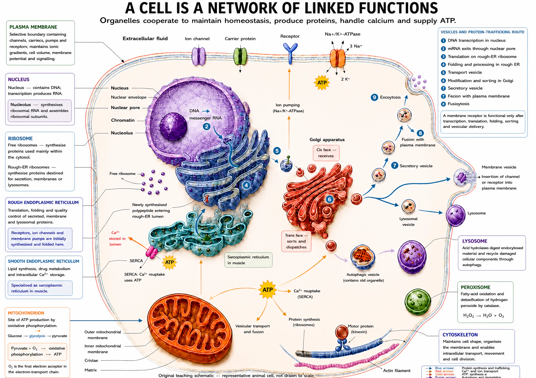
Two corrections to read alongside this diagram. Its numbered trafficking list mislabels the final steps — the correct order is 7 secretory vesicle → 8 fusion with the plasma membrane → 9 exocytosis, set out in full below. Its mitochondrion box also compresses ATP production to “pyruvate + O₂ → oxidative phosphorylation”; oxygen does not react with pyruvate directly. The real sequence is pyruvate → acetyl-CoA → TCA cycle → NADH/FADH₂ → electron-transport chain, with oxygen as the final electron acceptor and the proton gradient driving ATP synthase — developed in §03.
| Structure | Function | Anaesthetic relevance |
|---|---|---|
| Plasma membrane | Selective boundary containing transporters, channels, receptors and anchoring proteins. | Maintains ionic gradients, volume and excitability; a target for many anaesthetic drugs. |
| Nucleus and nucleolus | Deoxyribonucleic acid (DNA) storage/transcription; nucleolus makes ribosomal ribonucleic acid (rRNA) and assembles ribosomal subunits. | Allows longer-term adaptation by changing protein expression. |
| Ribosomes and rough endoplasmic reticulum (rough ER) | Translate and fold proteins for secretion, membranes or lysosomes. | Make receptors, channels, peptide hormones and secreted proteins. |
| Smooth endoplasmic reticulum (ER) / sarcoplasmic reticulum (SR) | Lipid synthesis, detoxification and calcium storage. | SR calcium release and re-uptake couple excitation to contraction. |
| Golgi apparatus and vesicles | Modify, sort and package proteins; vesicles deliver cargo or fuse for secretion. | Calcium-dependent exocytosis releases neurotransmitter. |
| Mitochondria | Citric-acid cycle, electron transport and oxidative phosphorylation. | ATP failure is central to hypoxia, ischaemia, shock and reperfusion injury. |
| Lysosomes and peroxisomes | Degradation/recycling; oxidative metabolism and peroxide handling. | Important in autophagy, cellular clean-up and oxidative injury. |
| Cytoskeleton and cell junctions | Shape, intracellular transport, movement, division and attachment to neighbouring cells. | Supports axonal transport and maintains membrane organisation; junctions compartmentalise tissues (e.g. tight junctions at the blood-brain barrier). |
The protein-trafficking route, in the order it actually happens
A membrane receptor, channel or secreted protein cannot affect cellular physiology until this full route has run. Some proteins (those staying in the cytosol) skip the rough-ER/Golgi portion entirely.
- 1 · DNA transcription in the nucleus produces messenger ribonucleic acid (mRNA).
- 2 · mRNA exits through a nuclear pore into the cytosol.
- 3 · Translation begins on a free cytosolic ribosome; a signal sequence in the growing chain directs the ribosome-mRNA complex to the rough endoplasmic reticulum (ER).
- 4 · Folding and initial processing occur inside the rough ER as translation continues.
- 5 · A transport vesicle buds from the ER and carries the folded protein to the Golgi apparatus.
- 6 · Golgi modification and sorting (for example glycosylation) address the protein to its final destination.
- 7 · A secretory vesicle buds from the Golgi’s trans face, carrying the protein toward the plasma membrane.
- 8 · The vesicle fuses with the plasma membrane.
- 9 · Exocytosis releases the cargo, or — for a channel or receptor — inserts the protein into the membrane itself, where it can finally do its physiological job.
Bioenergetics
ATP is the immediate energy currency
- Glycolysis in cytosol converts glucose to pyruvate and supplies a small ATP yield without directly requiring oxygen.
- With oxygen available, pyruvate enters mitochondria and becomes acetyl-coenzyme A (acetyl-CoA).
- The citric-acid (Krebs/TCA) cycle oxidises acetyl-CoA and generates the reduced electron carriers NADH and FADH₂, plus a small amount of ATP/GTP directly.
- The electron-transport chain in the inner mitochondrial membrane accepts electrons from NADH and FADH₂ and passes them down a series of carriers, pumping protons (H⁺) across the inner membrane and creating a proton electrochemical gradient. Oxygen is the final electron acceptor, combining with electrons and H⁺ to form water — it never reacts with pyruvate directly.
- The proton gradient drives protons back through ATP synthase, which phosphorylates ADP to ATP. This coupling of electron transport to phosphorylation is oxidative phosphorylation, and it is what oxygen delivery is actually for.
When oxygen supply is inadequate, pyruvate is converted to lactate instead of entering mitochondria, so glycolysis can continue regenerating the NAD⁺ it needs. This preserves a limited ATP supply but cannot replace mitochondrial oxidative phosphorylation in highly aerobic tissues such as brain and myocardium.
Where the ATP is spent
Membrane ion pumps — above all the Na⁺/K⁺-ATPase, which builds the sodium and potassium gradients Sections 05–07 depend on — are among the largest continuous ATP consumers in most cells, alongside protein synthesis, the sarco/endoplasmic reticulum Ca²⁺-ATPase (SERCA) and cytoskeletal/motor-protein work. This is why pump failure is the first functional consequence of ATP depletion, not a late or incidental one — the cell is already spending heavily on these pumps at rest.
With the energy budget established, the next three sections build the structure that budget maintains: the membrane itself, the transport mechanisms it hosts, and the water movement those mechanisms control.
Fluid mosaic model
The membrane is a fluid lipid bilayer studded with functional proteins
The plasma membrane — fluid mosaic model
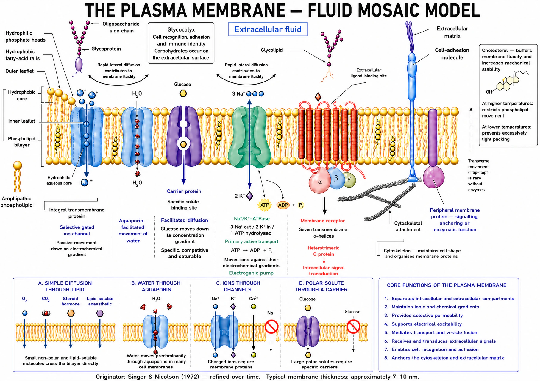
Phospholipids are amphipathic: a hydrophilic phosphate head faces water on either side, while two hydrophobic fatty-acid tails pack together to form the bilayer core, so the membrane is a barrier to water-soluble and charged solutes at the same time as it stays fluid enough for proteins to move within it.
| Component | Location | Structural role | Why it matters physiologically |
|---|---|---|---|
| Phospholipids | Both leaflets | Form the bilayer itself; tail saturation sets packing density | The barrier to water-soluble and charged solutes |
| Cholesterol | Intercalated among the tails, hydroxyl at the head region | Modulates fluidity and mechanical stability — buffers fluidity across a temperature range | Keeps the membrane functional whether cooled or warmed |
| Integral (transmembrane) proteins | Span the bilayer | Channels, carriers, pumps, receptors, enzymes | Do the work a lipid bilayer alone cannot |
| Peripheral proteins | Attached to one face only, usually cytoplasmic | Anchoring, signalling scaffolds, cytoskeletal linkage | Shape, mechanical support, localisation of signalling |
| Glycoproteins and glycolipids | Carbohydrate chains project into extracellular fluid only (the glycocalyx) | Cell recognition, adhesion, blood-group and immune identity | Restricted sidedness makes the membrane asymmetric, not just layered |
Cholesterol's effect on fluidity is temperature-dependent, and the direction matters. Above the membrane's phase-transition temperature, cholesterol's rigid ring system wedges between disordered phospholipid tails and reduces fluidity. Below that temperature, it disrupts the tight packing tails would otherwise adopt and increases fluidity — narrowing the temperature range over which membrane fluidity changes sharply, relevant across deliberate intraoperative hypothermia and fever alike.
Anaesthetic relevance — the Meyer–Overton correlation
Anaesthetic potency correlates closely with lipid solubility (the Meyer–Overton correlation) across chemically unrelated agents. This was historically read as evidence that anaesthetics act by dissolving in and disordering the bulk lipid bilayer. The correlation is real and durable, but bulk lipid disruption does not reproduce the anaesthetic state. Current evidence instead points to direct, saturable binding at discrete amphiphilic cavities within specific membrane proteins — Section 10 develops this in full.
Only small, lipid-soluble molecules and water cross the bilayer directly at a physiologically useful rate; everything else — ions, glucose, amino acids — depends entirely on the channel, carrier and pump proteins this structure hosts, which the next section works through in turn.
Moving solutes across the barrier
Every transport mechanism answers the same four questions differently
| Symbol | Meaning | Effect on diffusion |
|---|---|---|
| ṅ | Amount crossing per unit time | The result |
| P | Permeability (partition coefficient, diffusion coefficient, membrane thickness combined) | Greater P increases flux |
| A | Surface area available for exchange | Greater area increases flux |
| C₁ − C₂ | Concentration difference across the membrane | Greater gradient increases flux |
Simple diffusion needs no protein and does not saturate — flux keeps rising as the gradient steepens, unlike every carrier-mediated process below. Facilitated diffusion uses a carrier but is still passive: glucose moves down its electrochemical gradient through GLUT transporters without ATP. Because carrier number is finite, every carrier-mediated process shares three properties: specificity, saturation at a transport maximum (Tmax), and competition from structurally similar solutes.
Facilitated diffusion saturates; simple diffusion does not
A finite number of carrier proteins gives facilitated diffusion a transport maximum; simple diffusion through the lipid bilayer keeps rising as concentration rises. Axis values are illustrative (arbitrary units), not measured data.
| Protein | Physical mechanism | Key property | Rate behaviour | Example |
|---|---|---|---|---|
| Ion channel | Water-filled selective pore | Open probability/gating | High conductance when open | Voltage-gated Na⁺ channel, aquaporin |
| Carrier | Binds solute and alternately exposes its binding site | Specificity, competition, saturation | Finite transport maximum (Tmax) | Glucose transporter (GLUT) |
| Pump | Carrier coupled directly to a chemical energy source | Specificity and saturation | Moves solute against its gradient | Na⁺/K⁺-ATPase |
| Symporter | Couples two solutes moving in the same direction | Uses energy stored in a gradient | Secondary active | Na⁺–glucose cotransporter (SGLT) |
| Antiporter | Couples two solutes moving in opposite directions | Uses energy stored in a gradient | Secondary active | Na⁺/Ca²⁺ exchanger |
Primary active transport: the Na⁺/K⁺-ATPase builds the gradients everything else spends
The sodium–potassium adenosine triphosphatase moves three sodium ions out and two potassium ions in for each ATP hydrolysed. It is electrogenic (the unequal 3:2 exchange contributes a small direct current), but its dominant importance is maintaining the Na⁺/K⁺ gradients that cell-volume regulation, the resting membrane potential (Section 07) and secondary active transport all depend on. Sarco/endoplasmic reticulum Ca²⁺-ATPase (SERCA) and the plasma-membrane Ca²⁺-ATPase use the same primary-active principle to keep resting cytosolic calcium roughly four orders of magnitude below extracellular calcium — the gradient Section 09 relies on for calcium signalling.
Secondary active transport spends, rather than creates, a gradient. The sodium-glucose cotransporter (SGLT) uses the inward electrochemical pull on Na⁺ to drag glucose uphill — a symporter. The Na⁺/Ca²⁺ exchanger uses the same stored gradient to extrude calcium — an antiporter. Neither hydrolyses ATP itself; both stop working if the Na⁺/K⁺-ATPase is poisoned, which is why "active transport" is defined by movement against a gradient, not by which step consumes ATP.
| Mechanism | Carrier | Direction relative to gradient | Saturates | Energy source | Example |
|---|---|---|---|---|---|
| Simple diffusion | None | Down concentration gradient | No | No | O₂, CO₂, volatile anaesthetic agents, other lipid-soluble molecules |
| Channel-mediated diffusion | Ion channel | Down electrochemical gradient | Yes (gating), not saturation in the Michaelis–Menten sense | No | K⁺ leak channels, ligand-gated Na⁺ channels |
| Facilitated (carrier-mediated) diffusion | Carrier | Down electrochemical gradient | Yes — saturates at Tmax | No | GLUT-mediated glucose entry |
| Primary active transport | ATPase pump | Against electrochemical gradient | Yes — saturates | Direct (ATP hydrolysis) | Na⁺/K⁺-ATPase; sarco/endoplasmic reticulum Ca²⁺-ATPase (SERCA) |
| Secondary active transport | Cotransporter / exchanger | One solute downhill drives another uphill | Yes — saturates | Indirect (a pre-existing gradient) | Na⁺-glucose cotransporter (SGLT); Na⁺/Ca²⁺ exchange |
| Vesicular transport | Vesicle formation and membrane fusion | Bulk transfer of cargo too large for a channel or carrier | No | Cellular ATP-dependent machinery | Receptor-mediated endocytosis; regulated exocytosis |
Predict what happens if: extracellular glucose is pushed far above the physiological range — facilitated GLUT-mediated entry approaches its transport maximum and plateaus, unlike simple diffusion of a lipid-soluble gas, which keeps rising linearly with the gradient.
Water movement
Osmolality describes particles; tonicity predicts cell volume
Tonicity predicts the cell-volume response
Water moves toward the side with higher effective osmotic pressure. Labels assume a membrane that is poorly permeable to the effective extracellular solute.
Cell swells
Volume unchanged
Cell shrinks
| Term | Definition | Why it matters |
|---|---|---|
| Osmolarity | Osmoles of solute per litre of solution | Volume-dependent; the clinically estimated value |
| Osmolality | Osmoles of solute per kilogram of solvent | Mass-dependent — what a laboratory osmometer actually measures |
| Reflection coefficient, σ | How completely a membrane excludes a solute (0 = freely permeant, 1 = fully impermeant) | A non-penetrating solute exerts its full osmotic effect; a freely penetrating one exerts almost none |
| Tonicity | A solution's net effect on cell volume | Depends on effective osmoles only — a property of the solute-membrane pair, not of the solution alone |
Worked example — osmolarity vs osmolality, and why the distinction matters
150 mmol L⁻¹ NaCl (i = 2) plus 5 mmol L⁻¹ glucose (i = 1): osmolarity = (150 × 2) + (5 × 1) = 305 mOsm L⁻¹. In 1 L of water whose mass is 0.994 kg once solutes are dissolved, osmolality = 305 ÷ 0.994 ≈ 307 mOsm kg⁻¹ — close but not identical, because osmolarity is volume-based and osmolality is mass-based; the gap widens as solute concentration rises.
Hypotonic extracellular fluid drives net water entry and cell swelling — worsening cerebral oedema, which is why hypotonic intravenous fluids are avoided in neurological injury. Hypertonic fluid (hypertonic saline, mannitol) draws water out and is used to reduce intracranial pressure. With water movement and cell volume now explained by the same gradients the Na⁺/K⁺-ATPase builds, the next section turns those same gradients into a voltage.
From gradients to a voltage
Electrochemical gradients set the resting and action potential
| Gradient | Rule | Example |
|---|---|---|
| Chemical | Movement from higher to lower concentration | K⁺ tends to leave most cells |
| Electrical | Cations move toward negative potential; anions toward positive potential | Negative cell interior attracts cations |
| Electrochemical | Net effect of chemical and electrical forces | At rest, both forces drive Na⁺ inward |
The Nernst equation: one ion’s equilibrium potential
| Symbol | Meaning | Units |
|---|---|---|
| Eion | Equilibrium potential for one ion | mV |
| R, T, F | Gas constant, absolute temperature, Faraday constant | SI units |
| z | Valence (charge) | dimensionless |
| [ion]o/[ion]i | Extracellular/intracellular concentration | consistent units — the ratio is what matters |
Worked example — potassium
[K⁺]o = 4, [K⁺]i = 140 mmol L⁻¹, z = +1. EK = 61.5 × log₁₀(4/140) ≈ −95 mV. Sanity check: potassium's chemical gradient favours efflux, so the inside must be negative enough at equilibrium to hold that efflux back — a negative EK is the expected sign.
Goldman-Hodgkin-Katz: the actual membrane potential with several permeant ions
Chloride's terms are inverted because it is an anion. Use Nernst for one ion's equilibrium potential; use Goldman-Hodgkin-Katz for the actual membrane potential of a membrane permeable to several ions at once — the two answer different questions, and naming either without that distinction leaves it unclear which was being calculated.
Change permeability or extracellular potassium
This simplified Goldman-Hodgkin-Katz model uses representative concentrations. It illustrates direction and relative effect, not a universal value for every cell.
Representative resting conditions: potassium permeability dominates, so Vm lies near EK but not exactly at it.
The pump builds the gradients; permeability decides the instant voltage
Without the Na⁺/K⁺-ATPase, the concentration gradients Nernst and Goldman-Hodgkin-Katz depend on would run down within minutes. But at any single instant, Vm is set by which channels are open and how permeable the membrane currently is to each ion — not by the pump acting directly on voltage. This is why a channel-blocking drug or a change in extracellular K⁺ can alter Vm within milliseconds, while pump inhibition changes the gradients only gradually.
The action potential is regenerative channel behaviour
Nerve action potential: voltage, phases and refractory periods
Representative neuron, schematic timescale. Landmark voltages are the source texts' own stated values — rest −70 mV, firing level (threshold) −55 mV, peak +30 mV. The spike runs toward the sodium equilibrium potential (+60 mV) without reaching it; the undershoot runs toward the potassium equilibrium potential (≈ −95 mV, the value derived from the Nernst equation above) without reaching that either. The gradual rise before the firing level is the local (electrotonic) response to the stimulus; the steep regenerative upstroke only begins once threshold is crossed. The absolute refractory period spans almost the whole spike, the relative period the undershoot.
| Period | Mechanism | Functional result |
|---|---|---|
| Absolute | Na⁺ channels are inactivated, not merely closed | No second action potential can be generated, however strong the stimulus |
| Relative | Some Na⁺ channels have recovered, but K⁺ conductance remains elevated | A second action potential is possible, but only with a stronger-than-usual stimulus |
Conduction velocity is set by axial resistance (larger diameter is faster) and membrane properties (myelination is faster, via saltatory conduction between nodes of Ranvier). Local-anaesthetic Na⁺-channel block and demyelination both slow or stop conduction by reducing available regenerative current or increasing leak — not by changing the resting potential itself. With the membrane now able to generate and propagate a voltage signal, the next section turns to how a chemical signal — a receptor and its ligand — uses that same membrane to communicate.
First messenger to response
Receptor family predicts the speed and mechanism of signalling
| Family | Location | Mechanism | Effector | Onset | Duration | Example | Anaesthetic relevance |
|---|---|---|---|---|---|---|---|
| Ligand-gated ion channel | Plasma membrane | Ligand directly changes channel opening | Ion flux | Milliseconds | Brief | Nicotinic acetylcholine; GABA-A | Neuromuscular block; volatile/IV agents potentiate GABA-A |
| G-protein-coupled receptor | Plasma membrane | Heterotrimeric G protein modulates channel or enzyme | cAMP, IP₃/DAG, Ca²⁺ | Seconds to minutes | Seconds to minutes | Adrenergic, muscarinic, opioid | Opioids and α2 agonists act via Gi/o |
| Enzyme-linked receptor | Plasma membrane | Intrinsic/associated kinase phosphorylates targets | Phosphorylated proteins | Minutes to hours | Hours | Insulin receptor | Perioperative glycaemic control |
| Intracellular/nuclear receptor | Cytoplasm or nucleus | Ligand-receptor complex changes transcription | New mRNA/protein | Hours to days | Days | Glucocorticoid, thyroid hormone | Perioperative steroid cover; not the target of neuromuscular drugs |
Ligand-gated ion channels: the fastest family
Ligand-gated ion channel: structure and gating mechanism
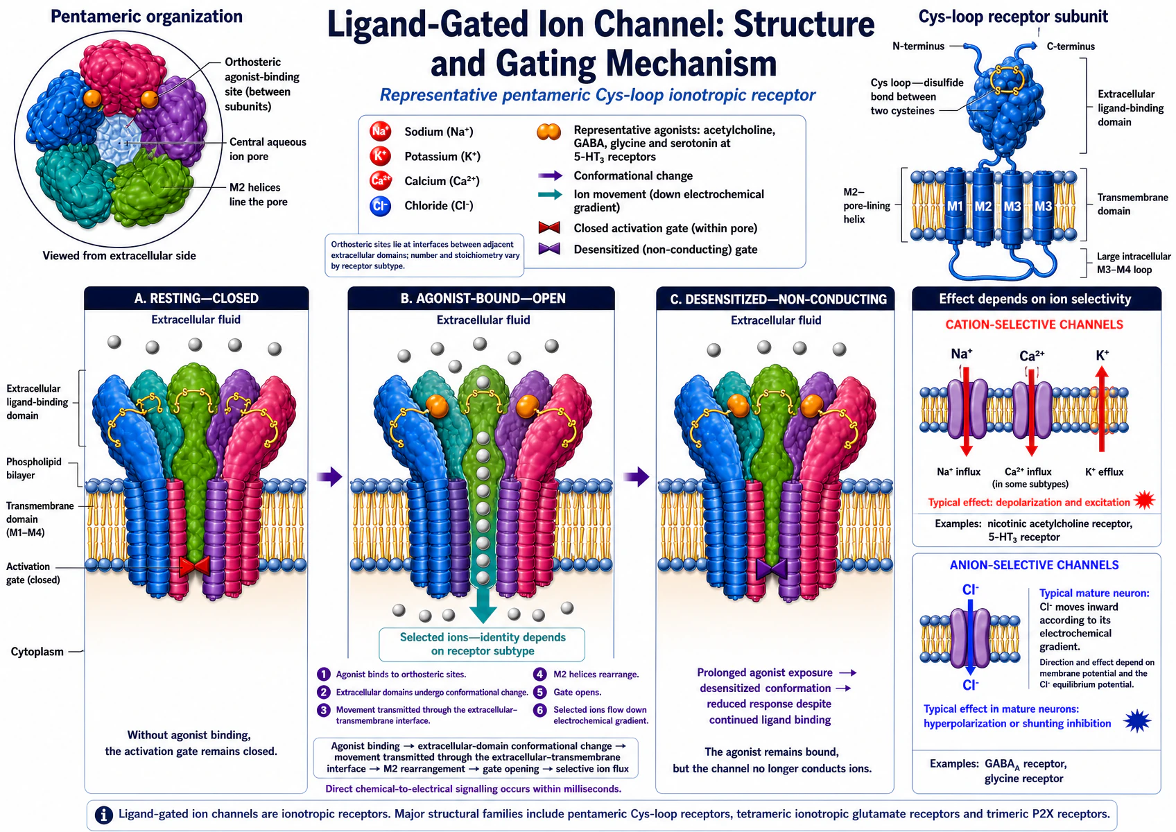
Effect depends on selectivity, not the receptor alone. Which ion moves, and which direction, depends on the pore's selectivity and the electrochemical gradient at the time. Chloride influx causing hyperpolarisation is the usual mature-neuron case (because mature neurons keep intracellular Cl⁻ low) — not a universal rule.
Worked example — the nicotinic receptor at the neuromuscular junction
Acetylcholine binds two α-subunits of the pentameric nicotinic receptor; the channel opens within microseconds and conducts both Na⁺ and K⁺, with net inward current because the driving force for Na⁺ entry is larger. This depolarises the end plate and, once threshold is crossed, triggers a propagating muscle action potential. Acetylcholinesterase terminates the signal within milliseconds — the pharmacological basis for both nondepolarising block (competitive antagonism) and its reversal with an anticholinesterase.
Intracellular and nuclear receptors: the slowest family
Type I and type II nuclear receptors
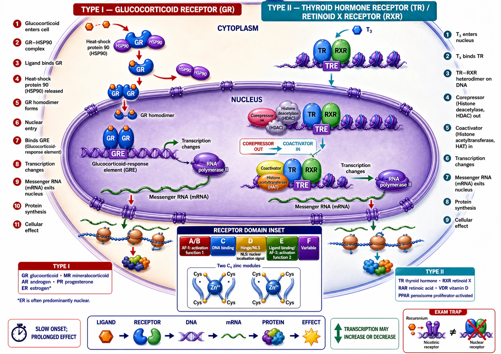
Not every type I receptor is cytosolic before ligand binds — this is the general pattern, not an absolute rule. The oestrogen receptor, formally type I, is predominantly nuclear even without ligand; state the mechanism for the specific receptor asked about.
| Type I (steroid-type) | Type II (thyroid-type) | |
|---|---|---|
| Resting location | Typically cytoplasmic, chaperoned by HSP90 | Typically already nuclear, bound to its DNA response element |
| On ligand binding | HSP90 released; receptor homodimerises and enters the nucleus | Corepressor exchanges for coactivator on the same DNA-bound complex |
| Examples | Glucocorticoid, mineralocorticoid, androgen, progesterone, oestrogen (predominantly nuclear even unliganded) | Thyroid hormone, retinoic acid, vitamin D, PPAR receptors |
Signal termination matters as much as generation: ligand removal, receptor desensitisation/internalisation, GTP hydrolysis, phosphatases and calcium re-sequestration all prevent an amplified response from becoming persistent noise — the G-protein branch of that termination machinery is next.
Amplification
G proteins couple a receptor to channels and enzymes
The heterotrimeric G-protein activation cycle
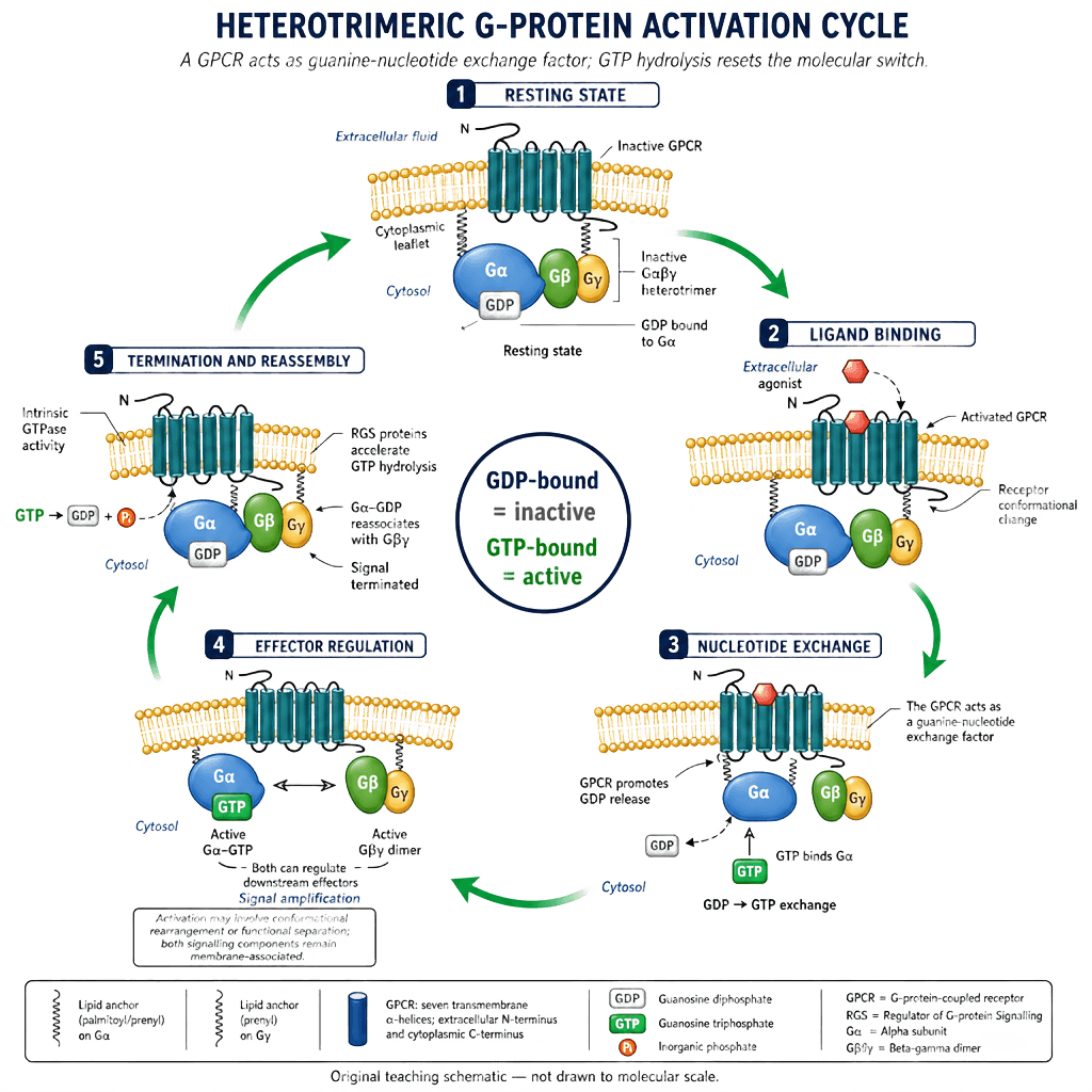
"Functionally separate" is more accurate than "dissociate." Both Gα-GTP and the freed Gβγ dimer stay membrane-anchored and can each regulate their own effector — activation is a conformational rearrangement, not necessarily complete physical separation. Gα's own intrinsic GTPase activity, accelerated by regulator-of-G-protein-signalling (RGS) proteins, makes the cycle self-terminating rather than a one-way switch.
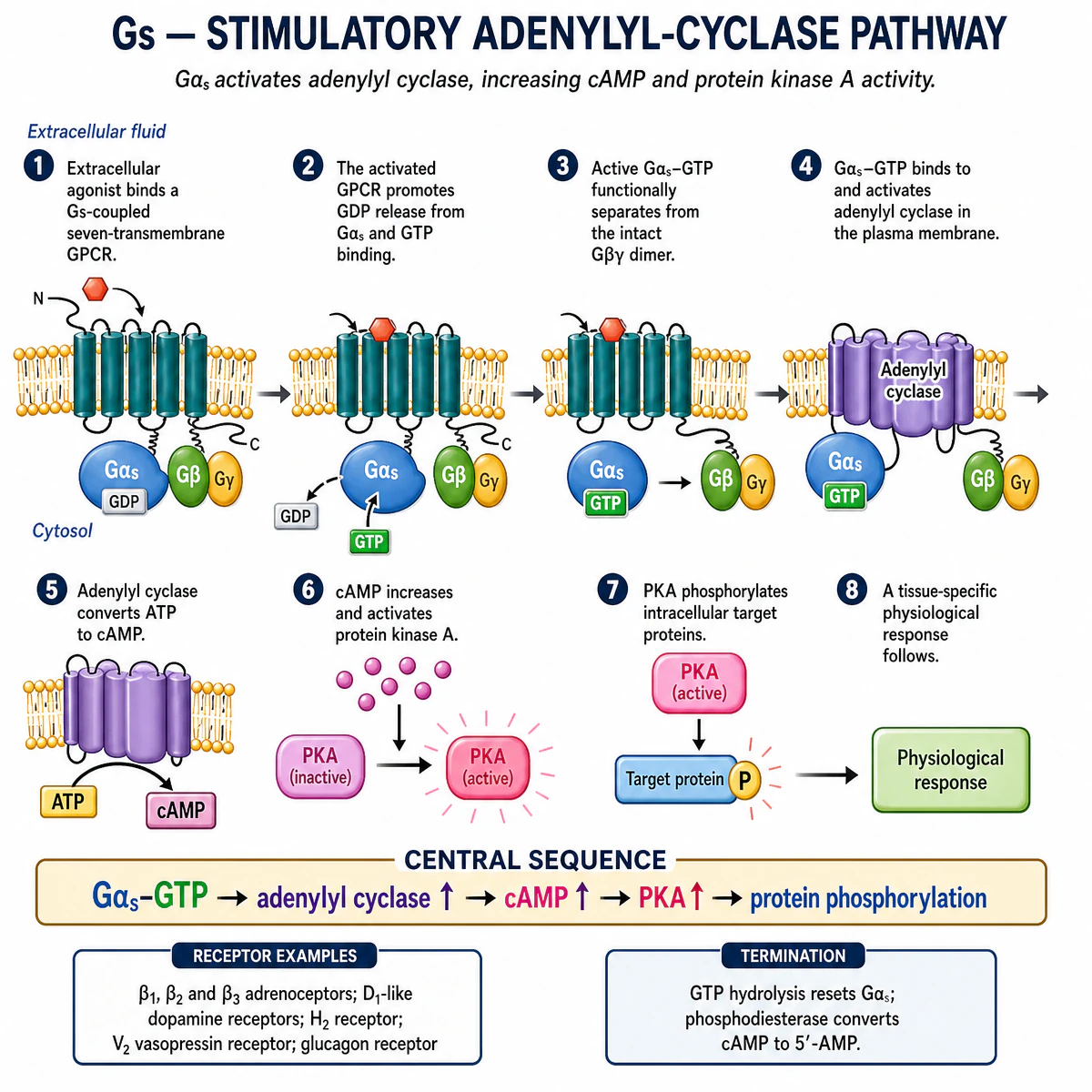
One receptor can activate many G proteins in turn, and each Gα-GTP or Gβγ can regulate its own effector — the basis of signal amplification, and the reason a handful of bound hormone molecules can produce a large, coordinated cellular response. Which effector responds depends on which Gα subtype is coupled to the receptor:
Gs — stimulatory adenylyl-cyclase pathway
Golf and Gt2 — specialised extension examples
Two further Gα subtypes use the same activation cycle for sensory transduction, beyond the core MMed Gs/Gi/Gq set above.
Golf — olfactory signal transduction
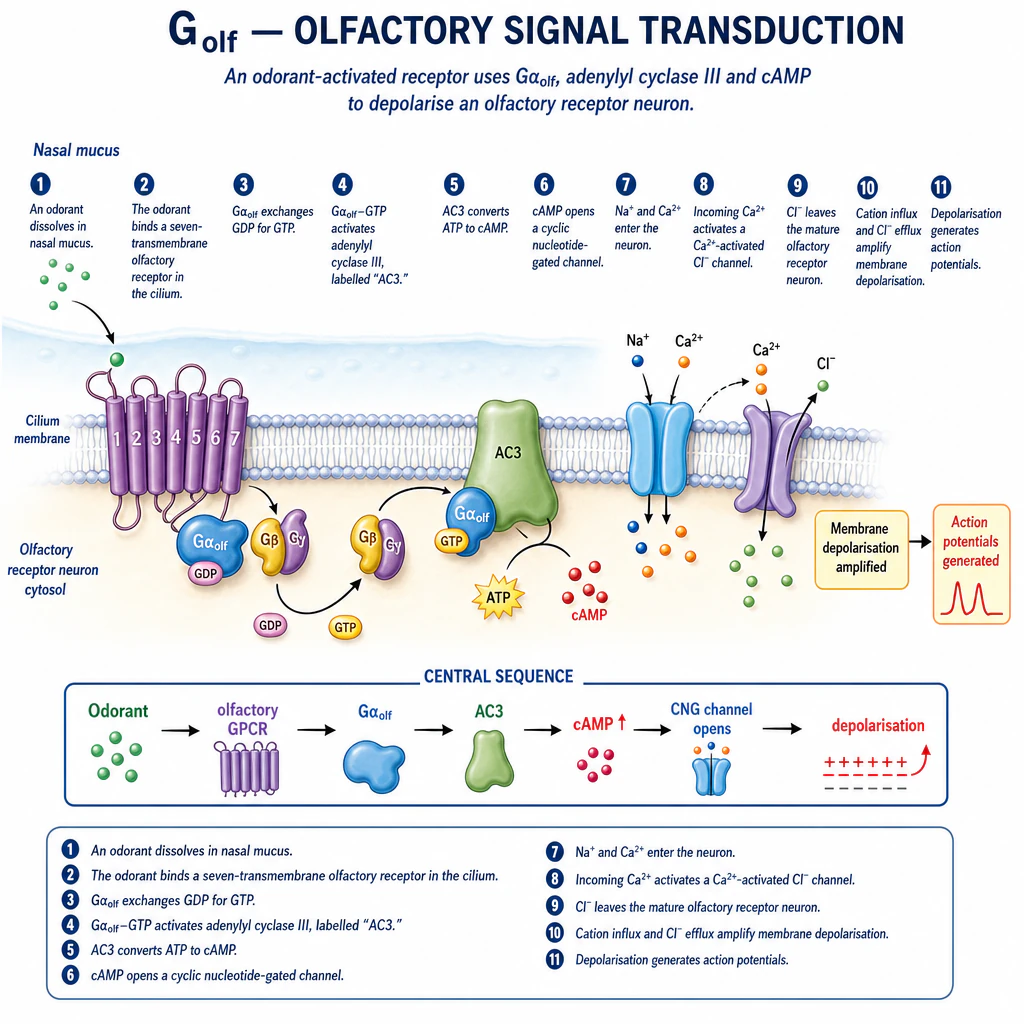
The chloride efflux shown reflects the mature olfactory-receptor-neuron chloride gradient specifically — the opposite direction to the usual mature-neuron GABA-A/glycine case, because these neurons hold intracellular Cl⁻ high enough for its efflux to be depolarising.
Gt2 (cone transducin) — phototransduction
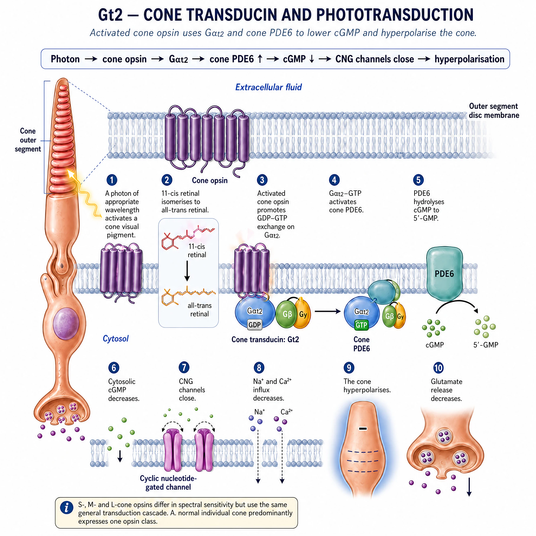
The opposite sign of change to Golf above — channel closure and hyperpolarisation, not opening and depolarisation — useful for showing the same GDP-GTP cycle can be coupled to either outcome depending on the effector.
Calcium is a signal only because resting calcium is low
Resting cytosolic free Ca²⁺ is held roughly four orders of magnitude below the extracellular concentration by continuous, energy-consuming work (Section 05): the plasma-membrane Ca²⁺-ATPase, the Na⁺/Ca²⁺ exchanger, and SERCA. Because the resting level is so low, a signal only has to open a channel — voltage-gated, or IP₃-/ryanodine-gated on the sarcoplasmic reticulum — to produce a large, rapid, easily detected relative rise. Calcium-dependent vesicle fusion is the final trigger for neurotransmitter release, and the same rise-and-clearance cycle underlies excitation-contraction coupling.
Predict what happens if: a Gs-coupled β1-adrenoceptor agonist and a phosphodiesterase inhibitor are given together. Both raise cAMP through different steps of the same pathway, so their positive inotropic and chronotropic effects would be expected to add.
Where the module closes the loop
Anaesthetic drugs act on these same proteins; losing this system is cellular injury
Meyer and Overton observed, around 1900, that anaesthetic potency correlates closely with lipid solubility across chemically unrelated agents (Section 04). This correlation is real and durable, but the mechanistic interpretation has changed: stereoselective differences between anaesthetic enantiomers of equal lipid solubility, together with direct structural evidence of anaesthetic binding within specific ion-channel proteins, now support direct protein binding rather than a bulk lipid-membrane effect.
| Target | Effect of anaesthetic binding | Functional consequence |
|---|---|---|
| GABA-A receptor (ligand-gated Cl⁻ channel) | Potentiation — increased channel open probability/duration at a given GABA concentration | Enhanced inhibitory synaptic transmission |
| Glycine receptor (ligand-gated Cl⁻ channel) | Potentiation, by a related mechanism to GABA-A | Enhanced inhibitory transmission, particularly spinal cord and brainstem |
| NMDA-type glutamate receptor | Inhibition (e.g. ketamine, nitrous oxide, xenon) | Reduced excitatory transmission — a mechanistically distinct effect from the GABAergic agents |
| Two-pore-domain (K2P) potassium channel | Direct activation | Increased background K⁺ conductance; hyperpolarises the resting membrane potential (Section 07) and reduces excitability |
These are not redundant descriptions of one effect: agents that potentiate GABA-A (most volatile agents, propofol, etomidate, benzodiazepines) and agents that inhibit NMDA instead (ketamine, nitrous oxide, xenon) produce recognisably different clinical anaesthetic states — evidence the two actions are mechanistically separate, not converging on one common pathway. No single receptor or theory fully explains general anaesthesia.
Immobility and unconsciousness are produced at different anatomical sites
Minimum alveolar concentration (MAC) — the concentration preventing movement in 50% of subjects at surgical incision — measures immobility specifically, generated largely at the spinal cord. Sedation, hypnosis and amnesia depend instead on supraspinal (cortical and subcortical) mechanisms. A technique that abolishes movement does not, by that fact alone, guarantee unconsciousness.
Cellular injury: the same system, failing
- Reduced oxygen delivery impairs oxidative phosphorylation (Section 03).
- ATP depletion impairs ion pumps and protein synthesis.
- Sodium and water entry (Section 05/06 in reverse) cause cell and organelle swelling; membrane potential collapses.
- Calcium accumulation (Section 09’s gradient, lost) activates phospholipases, proteases, endonucleases and ATPases.
- Mitochondrial and oxidative damage worsens ATP failure and injures membranes, proteins and DNA — reversible or irreversible depending on how far the sequence has run.
Hypoxia becomes cellular injury through adenosine triphosphate (ATP) depletion
A causal sequence. Severity, duration, substrate availability and reperfusion determine whether injury remains reversible.
- 1Reduced O₂ delivery
Hypoxaemia, low flow or impaired oxygen use.
- 2Oxidative phosphorylation falls
Less mitochondrial ATP.
- 3Pumps fail
Sodium–potassium pump (Na⁺/K⁺-ATPase) and calcium handling deteriorate.
- 4Na⁺ and water enter
Cell and organelles swell; membrane depolarises.
- 5Calcium overload
Proteases, phospholipases and endonucleases activate.
- 6Irreversible injury
Mitochondrial, membrane and DNA damage leads to necrosis or apoptosis.
| Feature | Reversible injury | Necrosis | Apoptosis |
|---|---|---|---|
| Morphology | Cell swelling and functional depression | Swelling, membrane rupture and contents leakage | Shrinkage, chromatin condensation and apoptotic bodies |
| Membrane | Sufficiently intact for recovery | Lost | Relatively maintained until phagocytosis |
| Inflammation | No primary response | Usually prominent | Usually limited |
| Meaning | Potential recovery if cause is reversed | Uncontrolled injury outcome | Controlled, energy-requiring cellular dismantling |
Autophagy — a cell recycling its own damaged organelles via the lysosome (Section 02) — is a survival response under moderate stress, distinct from both necrosis and apoptosis, and only worth invoking where it clarifies a specific question rather than as a third category to memorise alongside them.
Take-home points for this lesson. A cell spends energy to stay away from equilibrium; organelles build and deliver the proteins that do that work; oxidative phosphorylation pays for it and fails first when oxygen delivery fails; the Na⁺/K⁺-ATPase and its secondary transporters build the gradients that set water movement and membrane voltage; receptor family determines signalling speed; G proteins amplify a single ligand-binding event; general anaesthetics act through direct, saturable binding to a small number of confirmed protein targets; and cell injury is this entire system failing in a fixed causal order, with the point of no return set by how far that order has run.
Five ideas worth locking down
Five distinctions worth getting right
| The distinction | Where it is taught in full | Confused with |
|---|---|---|
| G proteins: the receptor is a nucleotide-exchange factor, not the effector | §09 G proteins | Naming Gs/Gi/Gq without explaining the GDP–GTP exchange cycle that activates them |
| Second messengers carry the signal onward from the G protein | §09 G proteins | Listing cAMP/IP₃/DAG/Ca²⁺ without saying which pathway produces which, or what each one then does |
| Glucose crosses membranes by facilitated diffusion, not active transport | §05 Transport | Classifying GLUT-mediated glucose transport as active, or an ATPase pump as facilitated diffusion |
| Resting membrane potential needs both the pump and permeability explained | §07 Electrical | Naming Nernst and Goldman-Hodgkin-Katz without explaining the principle behind either equation, or reversing the effect of extracellular K⁺ |
| Ionotropic vs metabotropic synapses are a named classification, not just "fast vs slow" | §08 Receptors | Treating "interneuron" or "saltatory conduction" as a type of synapse instead of the actual chemical/electrical, ionotropic/metabotropic classification |
Active recall: without looking back, state the six-step G-protein activation cycle, the Na⁺/K⁺-ATPase stoichiometry, the difference between Nernst and Goldman-Hodgkin-Katz, and the effect of hyperkalaemia on excitability. Retrieving them cold is what fixes them; if any comes out reversed or incomplete, that is the section to read again.