First messenger to response
Receptor family predicts the speed and mechanism of signalling
What you should already have
About 50 minutes
Plus the time it takes to redraw this lesson’s figures from memory, which is the fastest way to find out what you have not understood.
Where this shows up
Almost every drug given in an anaesthetic acts on a receptor described in this lesson, and the receptor family predicts how fast it will work and for how long. The second half is what happens when supply fails: the sequence from ATP depletion to cell death is the mechanism underneath every ischaemic injury seen in critical care.
Learning outcomes
By the end of this lesson you should be able to:
- Predict the speed and mechanism of signalling from the receptor family, and describe the structure and gating of a ligand-gated ion channel.
- Distinguish type I from type II nuclear receptors, and say why a nuclear receptor is not what rocuronium acts on.
- Describe the heterotrimeric G-protein activation cycle, and how a G protein couples a receptor to channels and enzymes.
- Explain why a rise in cytosolic calcium carries information only because resting calcium is held so low.
- Set out what is and is not established about the mechanism of general anaesthesia at the cellular level.
- Describe how hypoxia and ischaemia cause ATP depletion first and broad cellular dysfunction after it.
Together these settle one syllabus objective: Receptors, second messengers and cell injury. Tick it on the Physiology objective list once you can do all of the above without notes.
| Family | Mechanism | Typical speed | Example |
|---|---|---|---|
| Ligand-gated ion channel | Ligand directly changes channel opening | Milliseconds | Nicotinic acetylcholine; γ-aminobutyric acid type A (GABA-A) |
| G-protein-coupled receptor | Heterotrimeric G protein modulates channel or enzyme | Seconds to minutes | Adrenergic, muscarinic, opioid |
| Enzyme-linked receptor | Intrinsic/associated kinase phosphorylates targets | Minutes to hours | Insulin receptor |
| Cytokine receptor | Recruits kinases, often Janus kinase–signal transducer and activator of transcription (JAK-STAT) | Minutes to hours | Cytokine/growth-hormone receptors |
| Intracellular/nuclear receptor | Ligand-receptor complex changes transcription | Hours to days | Steroid, thyroid hormone |
Signal termination is physiologically important: ligand removal, receptor desensitisation/internalisation, GTP hydrolysis, phosphatases, phosphodiesterases and calcium re-sequestration prevent an amplified response from becoming persistent noise.
Ligand-gated ion channels: the fastest family
Ligand-gated ion channel: structure and gating mechanism
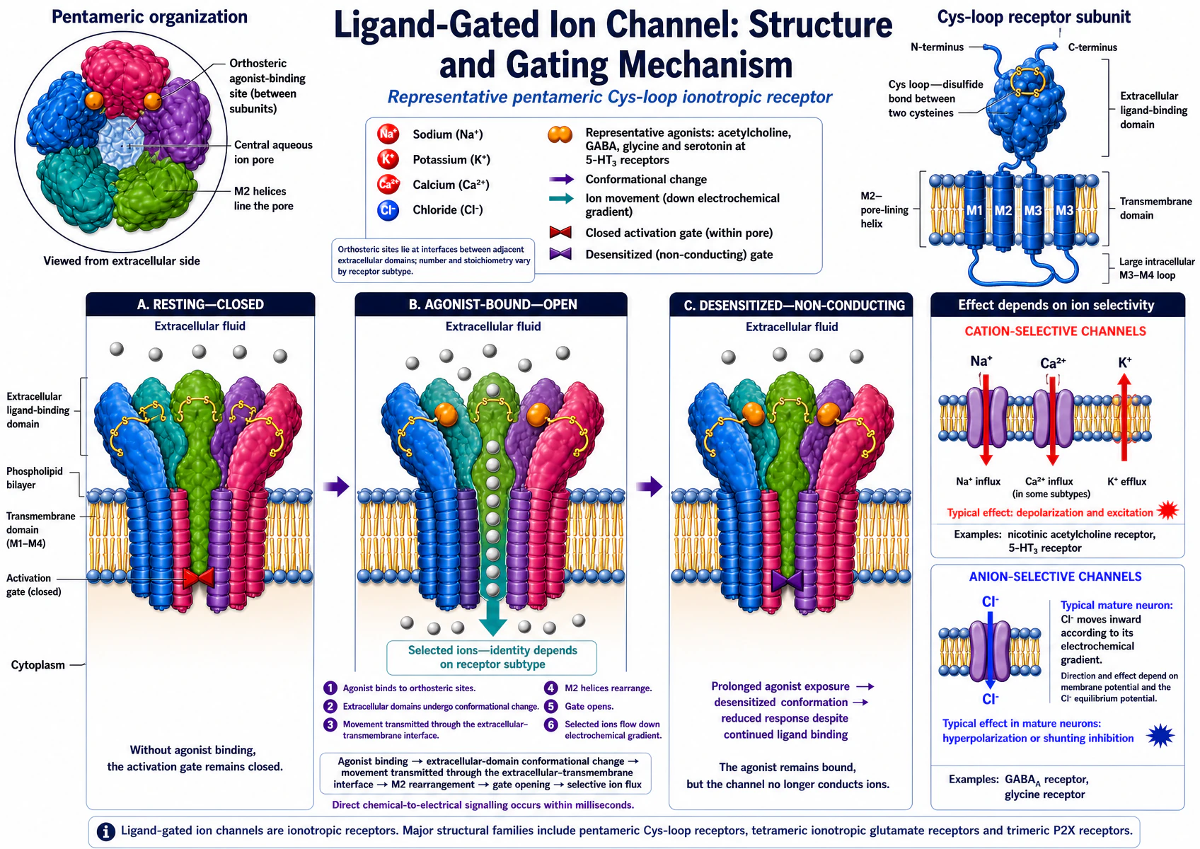
Effect depends on selectivity, not on the receptor alone. Which ion moves, and which direction, depends on the pore's ion selectivity and the electrochemical gradient at the time — not on ligand binding by itself. Chloride influx causing hyperpolarisation and inhibition is the usual case in a mature neuron, because mature neurons keep intracellular Cl⁻ low; it is not a universal rule (immature neurons, and cells with high intracellular Cl⁻, can show chloride efflux and depolarisation through the same GABA-A/glycine channels).
Worked example — the nicotinic receptor at the neuromuscular junction
The postjunctional nicotinic acetylcholine receptor (nAChR) is the clearest teaching example of a ligand-gated channel because its whole cycle is fast and stereotyped. Acetylcholine released from the nerve terminal binds two α-subunits of the pentameric receptor; the channel opens within microseconds and conducts both Na⁺ and K⁺, with net inward current because the driving force for Na⁺ entry is larger. This depolarises the end plate — the end-plate potential — and, once it exceeds the threshold for the surrounding sarcolemmal voltage-gated Na⁺ channels, triggers a propagating muscle action potential. Acetylcholinesterase in the synaptic cleft terminates the signal within milliseconds by hydrolysing acetylcholine, which is why neuromuscular block from a nondepolarising agent (competitive antagonism at the same binding site) or a depolarising agent (sustained occupancy that first opens, then desensitises, the channel) has such a fast onset and — for nondepolarising block — a reversal strategy built around out-competing the drug with a higher effective acetylcholine concentration (an anticholinesterase).
Intracellular and nuclear receptors: the slowest family
A lipid-soluble ligand (a steroid or thyroid hormone) crosses the plasma membrane directly and binds a receptor that itself changes gene transcription — hours to days slower than a channel or a GPCR, because the response depends on new protein synthesis. Type I and type II nuclear receptors differ in their resting location, not only in ligand.
Type I and type II nuclear receptors
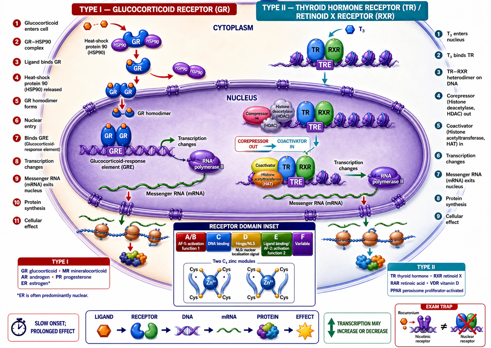
Not every type I receptor is cytosolic before ligand binds. This is the general pattern, not an absolute rule — the oestrogen receptor, formally type I, is predominantly nuclear even without ligand. State the mechanism for the specific receptor asked about rather than assuming every type I receptor is cytosolic.
| Type I (steroid-type) | Type II (thyroid-type) | |
|---|---|---|
| Resting location | Typically cytoplasmic, chaperoned by heat-shock protein 90 (HSP90) | Typically already nuclear, bound to its DNA response element |
| On ligand binding | HSP90 released; receptor forms a homodimer and enters the nucleus | Corepressor (with histone deacetylase) exchanges for coactivator (with histone acetyltransferase) on the same DNA-bound complex |
| DNA element | Hormone-response element (e.g. glucocorticoid-response element, GRE) | Response element bound as a heterodimer, usually with retinoid X receptor (RXR) |
| Examples | Glucocorticoid, mineralocorticoid, androgen, progesterone, oestrogen (predominantly nuclear even unliganded) receptors | Thyroid hormone, retinoic acid, vitamin D, peroxisome proliferator-activated (PPAR) receptors |
Ligand → receptor → DNA response element → messenger RNA (mRNA) → new protein → cellular effect. Transcription may be increased or decreased; the receptor and its bound coregulators set the direction, not just the fact, of the change.
Amplification
G proteins couple a receptor to channels and enzymes
The heterotrimeric G-protein activation cycle
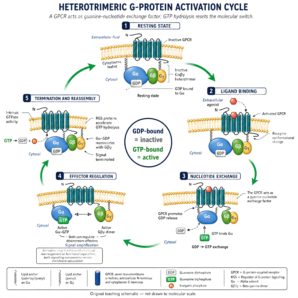
"Functionally separate" is more accurate than "dissociate." Both Gα-GTP and the freed Gβγ dimer stay membrane-anchored and can each regulate their own effector — activation is a conformational rearrangement, not necessarily complete physical separation. Gα's own intrinsic GTPase activity, accelerated by regulator-of-G-protein-signalling (RGS) proteins, is what makes the cycle self-terminating rather than a one-way switch.
Gs — stimulatory adenylyl-cyclase pathway
Golf and Gt2 — specialised extension examples
Two further Gα subtypes use the same activation cycle for sensory transduction, beyond the core MMed Gs/Gi/Gq set above.
Golf — olfactory signal transduction
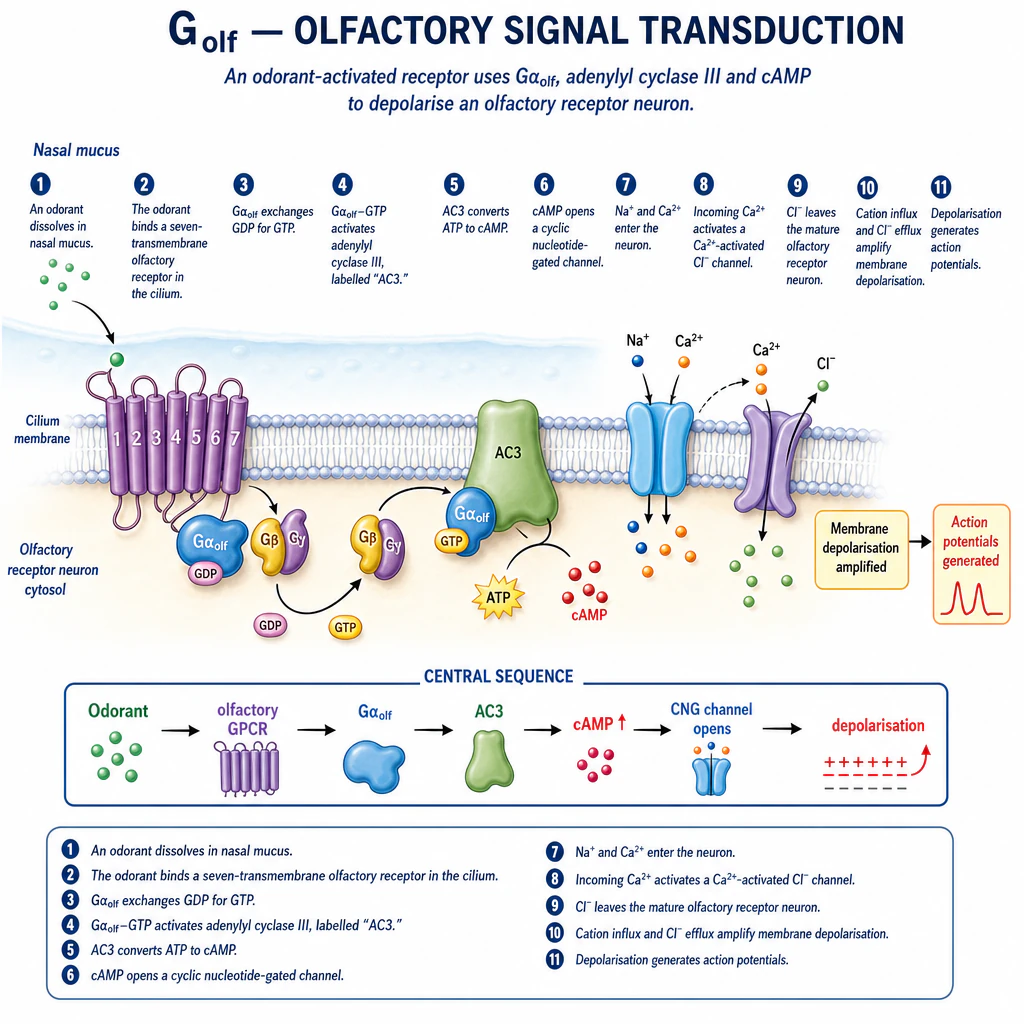
Gαolf couples odorant receptors to adenylyl cyclase III; the resulting cAMP rise opens a cyclic-nucleotide-gated cation channel. The chloride efflux shown reflects the mature olfactory-receptor-neuron chloride gradient specifically — the opposite direction to the usual mature-neuron GABA-A/glycine case above, because these neurons hold intracellular Cl⁻ high enough for its efflux to be depolarising rather than hyperpolarising.
Gt2 (cone transducin) — phototransduction
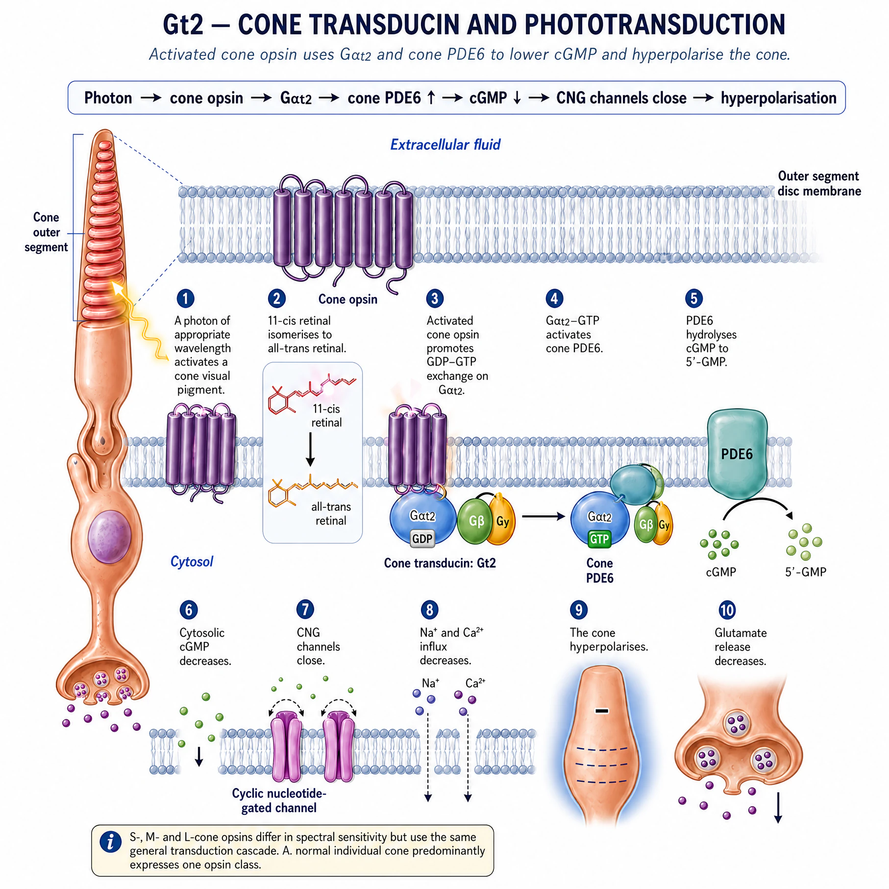
Transducin (Gt2) couples light-activated cone opsin to phosphodiesterase-6, which hydrolyses cGMP; falling cGMP closes cyclic-nucleotide-gated channels and hyperpolarises the cone, reducing glutamate release — the opposite sign of change to Golf above (channel closure and hyperpolarisation, not opening and depolarisation), useful for showing the same GDP-GTP cycle can be coupled to either outcome depending on the effector.
G proteins self-limit because the α-subunit’s own intrinsic GTPase activity hydrolyses GTP back to GDP — accelerated by regulator-of-G-protein-signalling (RGS) proteins — after which Gα-GDP reassociates with Gβγ. One receptor can activate many G proteins in turn, and each Gα-GTP or Gβγ can regulate its own effector: this is the basis of signal amplification — the reason a handful of bound hormone molecules can produce a large, coordinated cellular response.
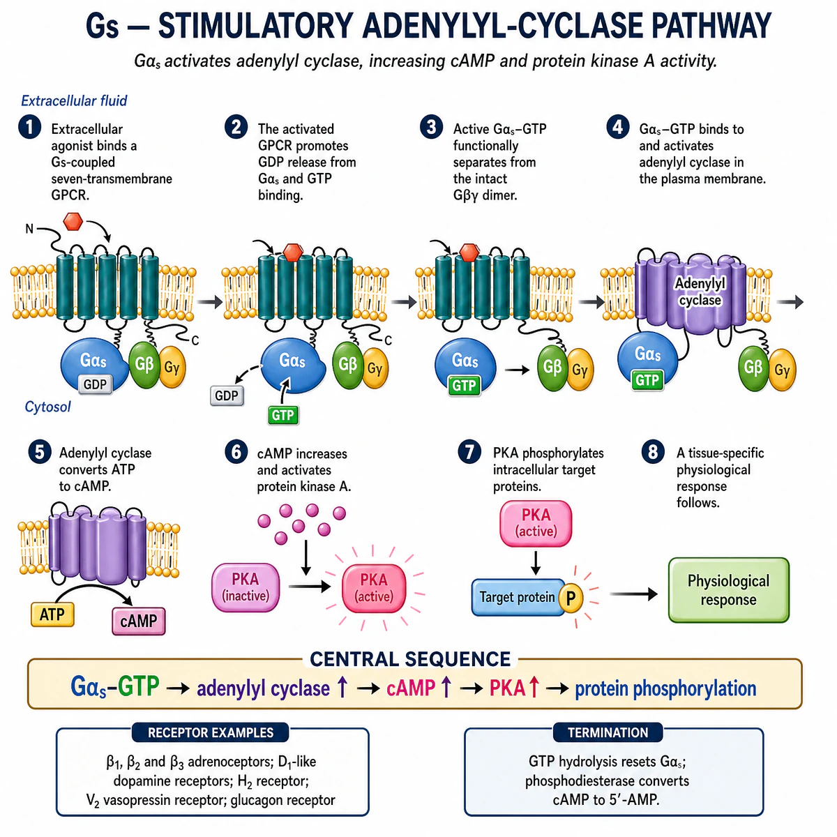
Predict what happens if: a Gs-coupled β1-adrenoceptor agonist and a phosphodiesterase inhibitor are given together. Both raise cAMP through different steps of the same pathway — the agonist by activating adenylyl cyclase, the inhibitor by slowing cAMP breakdown — so their positive inotropic and chronotropic effects would be expected to add.
A tightly controlled trigger
A rise in cytosolic calcium is useful only because resting calcium is low
Resting cytosolic free Ca²⁺ is held roughly four orders of magnitude below the extracellular concentration by continuous, energy-consuming work: the plasma-membrane Ca²⁺-ATPase and Na⁺/Ca²⁺ exchanger extrude calcium across the plasma membrane, and the sarco/endoplasmic reticulum Ca²⁺-ATPase (SERCA) pumps it back into internal stores. Because the resting level is so low, a signal only has to open a channel — a voltage-gated Ca²⁺ channel at the plasma membrane, or an IP₃- or ryanodine-gated channel on the sarcoplasmic reticulum — to produce a large, rapid, easily detected relative rise. Calcium-dependent vesicle fusion (via synaptotagmin sensing the local Ca²⁺ rise) is the final trigger for neurotransmitter and hormone release, and the same rise-and-clearance cycle underlies excitation-contraction coupling in cardiac and skeletal muscle.
Where anaesthetic drugs actually act
General anaesthetics bind directly to specific ligand- and voltage-gated channels
Meyer and Overton observed, around 1900, that anaesthetic potency correlates closely with a substance’s oil/water partition coefficient across chemically unrelated agents — the more lipid-soluble the drug, the more potent the anaesthetic. This correlation first focused a century of research on the lipid bilayer itself as the anaesthetic target. It has not been overturned as an empirical correlation, but the mechanistic interpretation has changed: stereoselective differences between anaesthetic enantiomers of equal lipid solubility, together with direct biophysical and structural evidence of anaesthetic binding within specific ion-channel proteins, now support direct protein binding rather than a bulk lipid-membrane effect as the operative mechanism.
| Target | Effect of anaesthetic binding | Functional consequence |
|---|---|---|
| GABA-A receptor (ligand-gated Cl⁻ channel) | Potentiation — increased channel open probability/duration at a given GABA concentration | Enhanced inhibitory synaptic transmission |
| Glycine receptor (ligand-gated Cl⁻ channel) | Potentiation, by a related mechanism to GABA-A | Enhanced inhibitory transmission, particularly in the spinal cord and brainstem |
| NMDA-type glutamate receptor | Inhibition (e.g. ketamine, nitrous oxide, xenon) | Reduced excitatory transmission; a distinct mechanism from the GABAergic agents |
| Two-pore-domain (K2P) potassium channel | Direct activation | Increased background K⁺ conductance; hyperpolarises the resting membrane potential and reduces excitability |
These are not redundant descriptions of one effect: agents that potentiate GABA-A receptors (most volatile agents, propofol, etomidate, benzodiazepines) and agents that inhibit NMDA receptors instead (ketamine, nitrous oxide, xenon) produce recognisably different clinical anaesthetic states, which is itself evidence that the two receptor actions are mechanistically separate rather than converging on one common final pathway.
Immobility and unconsciousness are produced at different anatomical sites
Minimum alveolar concentration (MAC) — the concentration that prevents movement in 50% of subjects in response to a surgical incision — is a measure of immobility specifically, not of unconsciousness. Spinal-cord transection and selective spinal-cord perfusion experiments show that the immobilising effect of an inhaled anaesthetic is generated largely at the level of the spinal cord, suppressing the motor response to a noxious stimulus, while sedation, hypnosis and amnesia depend instead on supraspinal (cortical and subcortical) mechanisms. This separation matters clinically: a technique that abolishes movement does not, by that fact alone, guarantee unconsciousness, which is part of the physiological basis for combining agents that act preferentially at different sites (for example, a volatile agent with an opioid) rather than relying on a single mechanism for every component of the anaesthetic state.
From reversible to irreversible
Hypoxia and ischaemia first cause ATP depletion, then broad cellular dysfunction
- Reduced oxygen delivery decreases oxidative phosphorylation.
- Adenosine triphosphate (ATP) depletion impairs ion pumps and protein synthesis.
- Sodium and water entry cause cell and organelle swelling; membrane potential collapses.
- Calcium accumulation activates phospholipases, proteases, endonucleases and ATPases.
- Mitochondrial and oxidative damage worsens ATP failure and injures membranes, proteins and deoxyribonucleic acid (DNA).
| Feature | Reversible injury | Necrosis | Apoptosis |
|---|---|---|---|
| Morphology | Cell swelling and functional depression | Swelling, membrane rupture and contents leakage | Shrinkage, chromatin condensation and apoptotic bodies |
| Membrane | Sufficiently intact for recovery | Lost | Relatively maintained until phagocytosis |
| Inflammation | No primary response | Usually prominent | Usually limited |
| Meaning | Potential recovery if cause is reversed | Uncontrolled injury outcome | Controlled cellular dismantling |
Membrane behaviour during injury connects back to membrane composition: a cooled or ischaemic membrane packs its phospholipid tails more tightly and cholesterol’s fluidising effect becomes proportionally more important, which is one reason therapeutic hypothermia — by slowing membrane-dependent enzyme kinetics and ATP consumption generally, not primarily through a fluidity effect — can extend the time available before ischaemic injury becomes irreversible.
Predict what happens if: reperfusion is delayed for a further ten minutes once ATP has fallen to a critically low level. Calcium continues to accumulate unchecked, phospholipase and protease activity continues, and the balance shifts further from reversible injury toward necrosis — this is why the duration of ischaemia, not only its presence, determines the clinical outcome.
Clinical integration
Cellular physiology predicts critical-care and anaesthetic consequences
| Principle | Clinical consequence |
|---|---|
| Hypotonic extracellular fluid | Water enters cells; cerebral cellular swelling may worsen |
| Hyperkalaemia | Resting depolarisation may progress to sodium-channel inactivation and impaired conduction |
| Local anaesthetic sodium-channel block | Action-potential propagation is slowed or blocked |
| Hypoxia/shock | ATP depletion impairs pumps, causes swelling and calcium overload |
| Reperfusion | Restored flow may also add reactive oxygen species and calcium-mediated injury |
| GABA-A potentiation / NMDA inhibition / K2P activation | The confirmed molecular targets through which general anaesthetic drugs alter cellular excitability |
| Malignant hyperthermia | Uncontrolled ryanodine-receptor calcium release triggered by volatile agents or succinylcholine in susceptible individuals |
Integrative mental model for the whole module
Three lessons, one connected system: the membrane’s lipid and protein composition (lesson 2) determines which transport mechanisms are physically possible; those transport mechanisms — above all the Na⁺/K⁺-ATPase — build and maintain the electrochemical gradients that set the resting and action potential (lesson 3); and the same channels and receptors that carry electrical signals also carry chemical ones, so a single set of proteins (GABA-A, glycine, NMDA, K2P, voltage-gated Na⁺ and Ca²⁺ channels) explains normal signalling, general anaesthetic action and the cellular consequences of hypoxia within one coherent framework, rather than as separate topics to memorise independently.
Common misconceptions
Take-home points for this lesson. Receptor family determines signalling speed and mechanism; G proteins amplify a single ligand-binding event into a large intracellular response; resting cytosolic calcium is kept low specifically so that a rise can act as a fast, detectable signal; general anaesthetic drugs act through direct, saturable binding to a small number of confirmed protein targets, with immobility and hypnosis generated at anatomically distinct sites; and cell injury follows a single causal chain from reduced oxygen delivery to ATP depletion to pump failure to calcium-mediated damage, with the point of no return set by how completely and for how long that chain has run.