Orientation
Rapid review — the whole topic in one panel
What you should already have
About 90 minutes
Plus the time it takes to redraw this lesson’s figures from memory, which is the fastest way to find out what you have not understood.
Where this shows up
Head position, carbon dioxide, oxygen, airway pressure, depth of anaesthesia and choice of agent all change intracranial pressure within minutes, and every one of them is in your hands. The physiology in this lesson is what makes the intervention right rather than habitual.
Learning outcomes
By the end of this lesson you should be able to:
- State the volume, formation rate and turnover of cerebrospinal fluid, and where it is produced.
- Describe the two-stage mechanism of secretion, and explain why composition proves it is secretion rather than filtration.
- Distinguish the blood-CSF barrier from the blood-brain barrier by the cell layer that forms each.
- Compare CSF with plasma for every measured constituent, and give the physiological consequence of each difference.
- List the functions of CSF, including its role as the first volume buffer for intracranial pressure.
- Trace the circulation from lateral ventricle to dural venous sinus, and explain why absorption is pressure-dependent while formation is not.
- Classify hydrocephalus physiologically and localise an obstruction from the pattern of ventricular dilatation.
Together these settle one syllabus objective: Cerebrospinal fluid: secretion, circulation, absorption and hydrocephalus. Tick it on the Physiology objective list once you can do all of the above without notes.
The minimum high-yield framework
- CSF — about 150 mL, made at about 0.35 mL min⁻¹ (≈ 500 mL day⁻¹), turning over three to four times daily. Mostly from the choroid plexus, by ultrafiltration then active secretion.
- Blood–CSF barrier — the choroid plexus epithelium, sealed by apical tight junctions. Its capillary is fenestrated on purpose.
- Composition — near-isosmotic with plasma, but lower potassium, lower glucose, higher chloride, far less protein, no cells, pH about 7.32.
- Functions — buoyancy, protection, a stable ionic environment, a volume buffer for ICP, acid–base signalling to the central chemoreceptors, and waste clearance.
- Route — lateral ventricles → foramina of Monro → third ventricle → cerebral aqueduct → fourth ventricle → foramina of Magendie and Luschka → cisterns and subarachnoid space → arachnoid villi → dural venous sinuses.
- Absorption is pressure-dependent; production is not. That asymmetry is the whole of CSF’s buffering capacity.
- ICP — 5–15 mmHg supine in an adult. Monro–Kellie doctrine: fixed total volume, so one compartment can only grow at another’s expense.
- Compliance = ΔV/ΔP; elastance = ΔP/ΔV. Reciprocals. The volume-pressure curve is flat, then knees, then steep.
- Compensation — CSF displacement and increased absorption first, venous blood second. Then nothing good is left.
- CPP = MAP − ICP. Autoregulation fails below a CPP of about 50 mmHg. The Cushing response is late.
The container
The intracranial compartments, and why the distinction between them matters
After the cranial sutures fuse, the adult cranial vault is a rigid container of fixed total volume. What is inside it is conventionally divided into brain tissue, blood and CSF; for planning what to do about a raised pressure it is more useful to split it four ways, because each has a different handle on it.
| Compartment | Share of intracranial volume | What it contains | Whether it can give way |
|---|---|---|---|
| Brain tissue | ≈ 80% (about 1400 g) | Neurons, glia and their intracellular fluid, plus brain interstitial fluid. | Not displaceable without injury. Grows with oedema, tumour, abscess or contusion. |
| Cerebrospinal fluid (CSF) | ≈ 10% (about 150 mL) | Ventricular and subarachnoid fluid, cranial and spinal. | The first and largest displaceable reserve — it can leave the cranium for the spinal subarachnoid space. |
| Blood — venous | part of ≈ 10% (about 150 mL of blood in total) | Dural venous sinuses and cerebral veins. | The second displaceable reserve. Passive, valveless and easily obstructed from outside the head. |
| Blood — arterial | part of the same ≈ 10% | Arteries and arterioles; cerebral blood volume is about 5 mL per 100 g of brain. | Not a reserve. Its volume is set by arteriolar tone, so it is the compartment anaesthesia changes fastest. |
The Monro–Kellie framework, stated early
Because the container is rigid, the sum of the compartment volumes is constant. An increase in any one of them must be matched by a decrease in one or both of the others, or intracranial pressure rises. That is the Monro–Kellie doctrine. Section 09 develops its pressure–volume consequences; here it is enough to notice its immediate corollary: only CSF and venous blood are genuinely displaceable, and both are small.
Definition and distribution
What CSF is, and where it is found
Where it is
- Ventricular CSF — the two lateral ventricles, the third ventricle, the cerebral aqueduct, the fourth ventricle and the central canal of the spinal cord. These surfaces are lined by ependyma, the ciliated epithelium modified over the choroid plexus into secretory epithelium.
- Cranial subarachnoid space — between arachnoid mater and pia mater, over the cerebral hemispheres, crossed by arachnoid trabeculae which help suspend the brain.
- The cisterns — the widened parts of the subarachnoid space at the skull base: cerebellomedullary (cisterna magna), prepontine, interpeduncular, ambient and quadrigeminal. Their obliteration on imaging is one of the recognised signs of a raised ICP or an exhausted compensatory reserve.
- Spinal subarachnoid space — continuous with the cranial space and extending to the lumbar cistern. This continuity is what makes CSF displacement possible, and therefore what gives the intracranial space any compensatory reserve at all.
Relationship to brain extracellular fluid
CSF and brain interstitial fluid communicate freely across the ependyma and the pial surface, and their compositions are essentially the same. That is why CSF sampled at the lumbar cistern tells you something about the brain’s chemical environment, and why the acid–base status of CSF is meaningful for a chemoreceptor sitting in the medulla rather than in the ventricle.
Secretion, not filtration
CSF production at the choroid plexus
CSF production at the choroid plexus
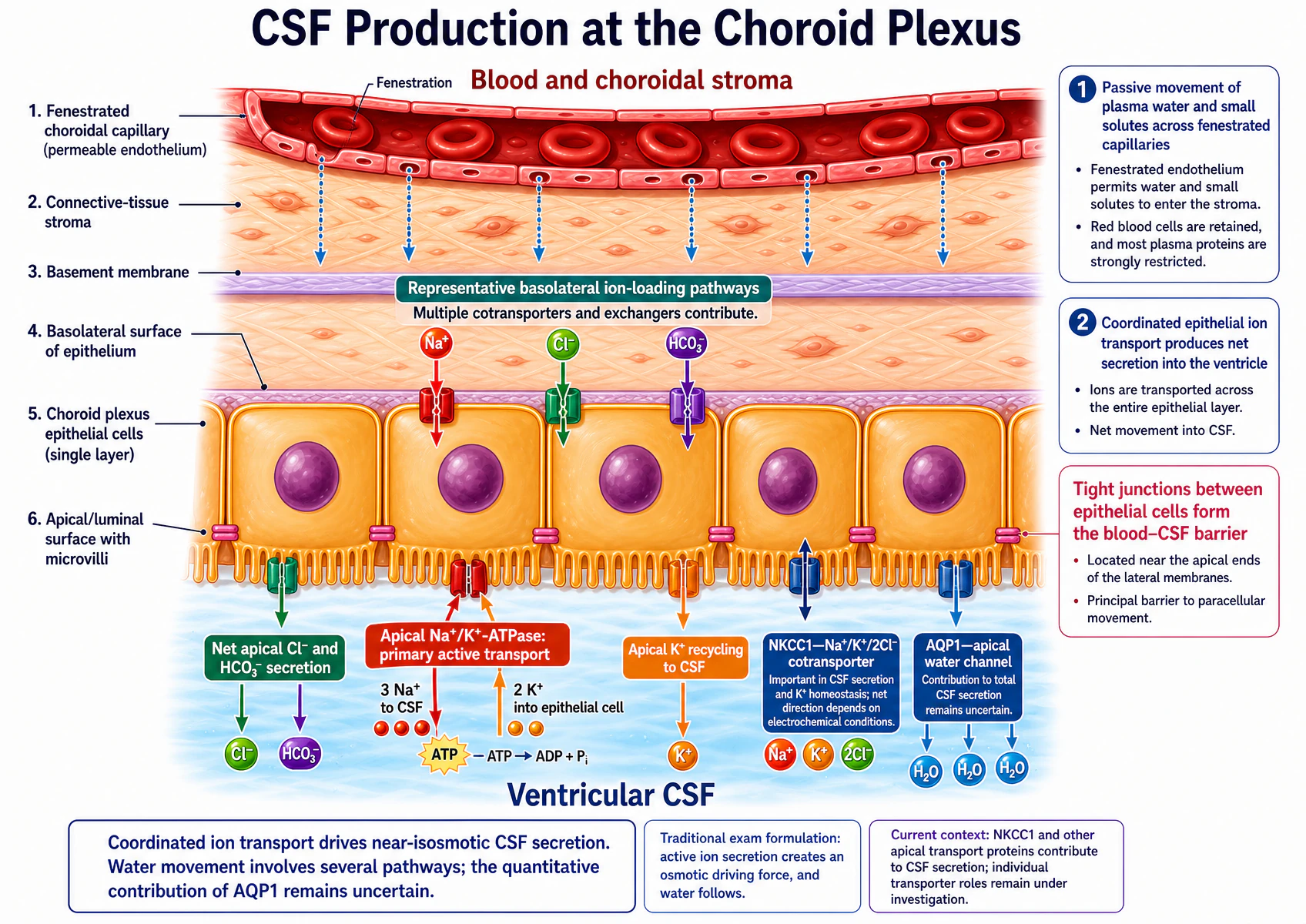
One qualification to read alongside this diagram. The choroid plexus epithelium is unusual among secretory epithelia in siting its Na⁺/K⁺-ATPase on the apical (CSF-facing) membrane, exactly as drawn — which is what allows the pump to extrude sodium directly into the ventricle and so drive net secretion. Some anaesthetic texts still place the pump on the basolateral membrane instead. The physiology does not depend on resolving that: the load-bearing point is that active, ATP-dependent sodium transport into the ventricle creates the osmotic gradient, and water follows. The basolateral arrows are labelled representative on the diagram itself for the same reason — several cotransporters and exchangers load the cell, and the figure shows the principle rather than a complete transporter inventory.
Stage 1 — ultrafiltration into the stroma
The choroidal capillary is fenestrated. This is deliberate and is the opposite of the arrangement in the rest of the brain: hydrostatic pressure drives plasma water and small solutes through the fenestrations into the connective-tissue stroma of the villus. Red cells are retained and most plasma protein is strongly restricted, so what reaches the stroma is a protein-poor ultrafiltrate. Nothing about this step is selective enough to explain CSF composition.
Stage 2 — active secretion across the epithelium
The selectivity comes from the single layer of cuboidal choroid plexus epithelium sitting on its basement membrane. Its cells are joined near their apical ends by tight junctions, and it is those junctions — not the capillary — that form the blood–CSF barrier and block the paracellular route. Solute therefore has to go through the cells, which is what makes the composition controllable.
- Basolateral loading. Sodium, chloride and bicarbonate are taken up across the basolateral surface by a set of cotransporters and exchangers. Bicarbonate and hydrogen ion are also generated inside the cell from carbon dioxide and water, a reaction catalysed by carbonic anhydrase.
- Apical extrusion of sodium. The apical Na⁺/K⁺-ATPase hydrolyses ATP to move three sodium ions out into the CSF and two potassium ions into the cell. This is the primary active step, and it is what keeps intracellular sodium low so that basolateral loading can continue.
- Anions follow. Chloride and bicarbonate move down the resulting electrochemical gradient into the CSF, so that the secreted fluid is electrically neutral.
- Potassium recycles. Potassium brought into the cell by the pump returns to the CSF through apical channels — which is part of why CSF potassium is held at a value well below plasma rather than simply rising with the pump.
- Water follows osmotically, through the cells (including apical aquaporin-1 channels) and to some extent between them, producing a fluid that is near-isosmotic with plasma — which is why CSF osmolality matches plasma even though its ionic composition does not.
How much, how fast, and from where
- Rate: about 0.35 mL min⁻¹ (texts give 0.3–0.4), which is ≈ 500 mL day⁻¹. Against a total volume of about 150 mL, that is a turnover of three to four times daily.
- Site: mostly the choroid plexus of the lateral, third and fourth ventricles. Estimates of its share range from about half to about 70%; the remainder comes from cerebral capillary endothelium and from water generated by brain metabolism.
- Regulation: production is essentially independent of intracranial pressure across the range that matters clinically. It falls only when cerebral perfusion pressure drops below about 70 mmHg and choroid plexus blood flow falls with it.
Why the numbers matter
CSF compared with plasma
| Constituent | CSF | Plasma | Why the difference matters |
|---|---|---|---|
| Sodium (Na⁺) | ≈ 140 mmol L⁻¹ | ≈ 140 mmol L⁻¹ | Essentially the same — CSF is near-isosmotic with plasma, not dilute. |
| Potassium (K⁺) | ≈ 2.9 mmol L⁻¹ | ≈ 4.5 mmol L⁻¹ | Lower, and tightly held there. Neuronal excitability is protected from plasma potassium swings. |
| Chloride (Cl⁻) | ≈ 120–125 mmol L⁻¹ | ≈ 100–110 mmol L⁻¹ | Higher in CSF — the anion that accompanies secreted sodium. |
| Bicarbonate (HCO₃⁻) | ≈ 21–25 mmol L⁻¹ | ≈ 23–25 mmol L⁻¹ | Similar. Slow bicarbonate movement is what makes CSF pH adapt over hours, not minutes. |
| Glucose | ≈ two-thirds of plasma | — | Facilitated transport, saturable. A markedly low CSF glucose suggests bacterial or tuberculous meningitis. |
| Calcium (Ca²⁺) and magnesium (Mg²⁺) | Calcium lower; magnesium higher | — | Both actively controlled, both matter for excitability. Texts differ on the exact ratios. |
| Protein | ≈ 0.2–0.4 g L⁻¹ | ≈ 70 g L⁻¹ | About 200-fold lower. Two consequences: almost no oncotic pressure, and almost no non-bicarbonate buffering. |
| Cells | 0–5 mm⁻³ | 4000–11 000 mm⁻³ (white cells) | Normally acellular. Any significant cell count is pathological. |
| pH | ≈ 7.32 | ≈ 7.40 | CSF is more acidic, and its carbon dioxide tension is higher than arterial. |
| Osmolality | ≈ 289 mOsm kg⁻¹ | ≈ 289 mOsm kg⁻¹ | Matched. Secretion is near-isosmotic; water follows the ions rather than being pumped. |
Four consequences worth stating out loud
- Neuronal excitability is protected. Potassium, calcium and magnesium set membrane potential and firing threshold. Because they are actively controlled at the choroid plexus rather than tracking plasma, a patient’s plasma potassium can move considerably without altering neuronal behaviour.
- CSF buffers weakly. Almost no protein means almost no non-bicarbonate buffer. So a change in carbon dioxide produces a larger swing in CSF pH than the same change produces in blood.
- Arterial carbon dioxide reaches the chemoreceptors quickly. Carbon dioxide crosses the blood–brain barrier freely; hydrogen ion and bicarbonate do not. So an acute rise in arterial carbon dioxide acidifies CSF within minutes and stimulates the central chemoreceptors, whereas a metabolic acidosis of the same severity does not — the barrier keeps the hydrogen ions out. This is why respiratory acidosis is the dominant acute ventilatory drive.
- Almost no oncotic pressure. With CSF protein about 200-fold lower than plasma, CSF oncotic pressure is effectively zero. Absorption at the arachnoid villi is therefore a straightforward hydrostatic bulk flow rather than a Starling balance — which is exactly why it is pressure-dependent (§06).
Mechanism, not a list
The functions of CSF
| Function | Mechanism | Physiological result | Anaesthetic or clinical relevance |
|---|---|---|---|
| Buoyancy | CSF has almost the same specific gravity as brain, so the brain floats in it. | Effective brain weight falls from about 1400 g to under 50 g, reducing inertia and traction on vessels and nerve roots. | Loss of CSF at lumbar puncture lets the brain sag on those attachments — the mechanism of post-dural-puncture headache. |
| Mechanical protection | A fluid layer between brain and the ridged skull base. | Damps acceleration and deceleration forces. | Explains why a blow to the head can injure the brain at the opposite pole (contrecoup). |
| A stable ionic environment | Active transport at the choroid plexus sets CSF composition independently of plasma. | Potassium, calcium and magnesium are held constant, so excitability is protected from plasma swings. | A patient's plasma potassium can move considerably without changing neuronal behaviour. |
| Volume buffer for intracranial pressure | CSF can leave the cranium for the spinal subarachnoid space, and its absorption rises with pressure. | The first compensation for any expanding intracranial mass, and the reason the volume-pressure curve has a flat limb at all. | Draining a few millilitres through a ventricular drain can drop a raised ICP substantially. |
| Acid–base signalling for respiratory control | Carbon dioxide crosses the blood–brain barrier freely; hydrogen ion and bicarbonate do not. | CSF pH tracks arterial carbon dioxide closely, and weak CSF buffering makes that response large and fast — the signal the central chemoreceptors read. | This is why arterial carbon dioxide, not arterial pH, is the dominant acute drive to ventilation. |
| Distribution of signalling substances | Neuropeptides secreted into CSF are carried between brain regions. | A slow volume-transmission route alongside synaptic transmission. | One reason intrathecal drug administration reaches targets that intravenous administration cannot. |
| Waste clearance (glymphatic exchange) | CSF enters periarterial spaces, crosses the parenchyma through astrocyte aquaporin channels, and leaves by perivenous routes. | Substitutes for the lymphatic drainage the brain parenchyma lacks. | Periarterial spaces widen during sleep and general anaesthesia, so clearance rises; volatile agents reduce it more than dexmedetomidine. |
One fixed route
CSF circulation and absorption
CSF production, circulation and absorption
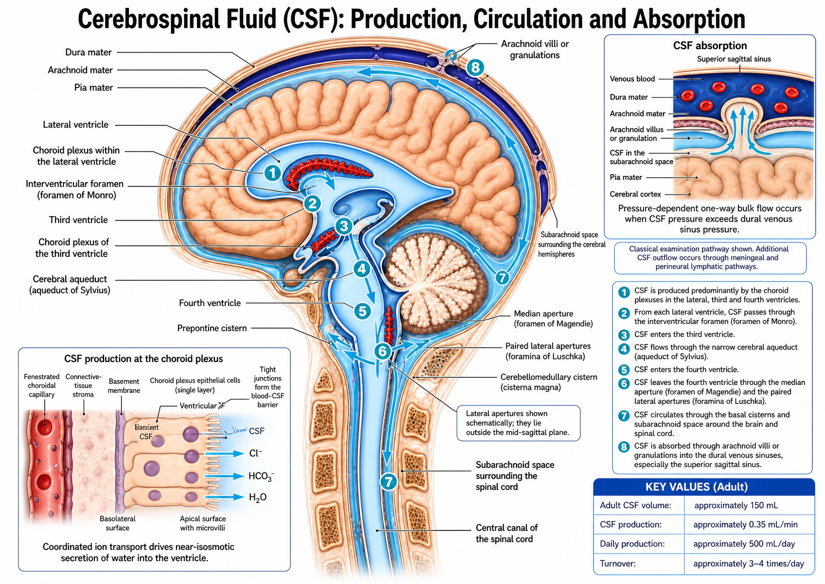
Read the lower-left production inset with these corrections. Three of its labels are damaged in the supplied artwork: the leader labelled “Ventricular CSF” is overprinted and partly illegible, and it points into the epithelial layer rather than into the ventricular lumen it names; a garbled overprinted fragment sits on top of the epithelial cells and is not a real label — ignore it; and the topmost of the four apical arrows, marked CSF, is drawn as a scribble rather than as a clean arrow like the three below it. The physiology the inset is trying to show is correct and is set out in full in §03. Note also that the inset shows only chloride, bicarbonate and water crossing the apical surface — sodium is the principal secreted cation and is shown properly in the choroid plexus diagram in §03.
Two labels on the main figure are qualifications rather than errors, and the artwork states both itself: the paired lateral apertures (foramina of Luschka) are drawn schematically because they do not lie in the mid-sagittal plane, and the arachnoid-villus route is presented as the classical examination pathway with additional meningeal and perineural lymphatic outflow noted alongside it. Both are correct as drawn. The key values — 150 mL, 0.35 mL min⁻¹, 500 mL day⁻¹, turnover three to four times daily — were each checked and are internally consistent with one another.
The ordered route, as you should be able to recite it
- Lateral ventricles — the largest choroid plexus, and where most CSF is made.
- Interventricular foramina (foramina of Monro), one from each lateral ventricle.
- Third ventricle, between the thalami, with its own choroid plexus.
- Cerebral aqueduct (aqueduct of Sylvius) through the midbrain — the narrowest point on the whole route, and therefore the commonest site of congenital obstruction.
- Fourth ventricle, between pons and cerebellum.
- Median aperture (foramen of Magendie) and the paired lateral apertures (foramina of Luschka) — the exit from the ventricular system into the subarachnoid space.
- Basal cisterns — cerebellomedullary (cisterna magna), prepontine, interpeduncular and the rest.
- Cranial and spinal subarachnoid space — most CSF passes over the hemispheres, the remainder around the cord.
- Arachnoid villi and granulations into the dural venous sinuses, chiefly the superior sagittal sinus.
Flow is driven by the pressure gradient from the site of production to the site of absorption, and assisted by ciliary action of the ependyma and by arterial pulsation of the cerebral vessels and choroid plexus, together with respiratory oscillation.
Absorption is pressure-dependent — and that is the important half
CSF is absorbed through arachnoid villi and granulations, outpouchings of arachnoid that project through the dura into the venous sinuses. Roughly 85–90% through intracranial villi (mainly of the sagittal and sigmoid sinuses) and 10–15% through spinal arachnoid villi around the dorsal nerve roots.
They behave as one-way, pressure-sensitive valves. Bulk flow into the sinus begins once CSF pressure exceeds dural venous sinus pressure by of the order of 1.5 mmHg, and thereafter absorption increases essentially linearly with CSF pressure. Below roughly 5–7 mmHg, absorption is negligible.
| Symbol | Meaning | Units | Interpretation |
|---|---|---|---|
| PCSF | CSF pressure in the subarachnoid space | mmHg (or cmH₂O) | Rises with any expanding intracranial volume |
| Psinus | Pressure in the dural venous sinus | mmHg (or cmH₂O) | Rises with central venous pressure, jugular obstruction or sinus thrombosis |
| Gradient | The difference between them | mmHg | Of the order of 4–5 mmHg normally; absorption stops when it closes |
This produces a genuine negative feedback: a rise in ICP raises CSF pressure, which increases absorption, which limits the rise. It is why a modest added volume can be absorbed away rather than converted into pressure. Two things break it — a rise in dural venous sinus pressure, which closes the gradient, and blocked granulations after subarachnoid haemorrhage or meningitis, which raise outflow resistance.
Applied CSF physiology
Hydrocephalus, and localising an obstruction from the imaging
The consequence is a rise in CSF pressure proximal to the problem, compression of brain parenchyma and dilatation of the affected ventricles — ventriculomegaly. Sustained compression causes irreversible damage, which is why the physiological classification matters: it tells you where to intervene.
| Type | Mechanism | Typical causes | Which parts dilate | Principle of management |
|---|---|---|---|---|
| Obstructive (non-communicating) | Block inside the ventricular system, before CSF reaches the subarachnoid space | Aqueduct stenosis; tumour at a foramen of Monro; Arnold–Chiari malformation obstructing the fourth-ventricle outlets | Only the parts proximal to the block | Relieve or bypass the obstruction — for example endoscopic third ventriculostomy for aqueduct stenosis |
| Communicating | CSF reaches the subarachnoid space but absorption fails at the arachnoid granulations | Blood clot after subarachnoid haemorrhage; chronic meningitis | The whole ventricular system | Divert CSF — external ventricular drain acutely, ventricular shunt for the longer term |
| Overproduction (rare) | No obstruction at all; formation simply exceeds absorptive capacity | Choroid plexus papilloma | The whole ventricular system | Remove the lesion |
Worked example — reading the site of obstruction off the scan
A child with congenital aqueduct stenosis. Work upstream and downstream of the block.
The cerebral aqueduct connects the third ventricle to the fourth. Upstream of it lie the third ventricle and, through the foramina of Monro, both lateral ventricles — so all three dilate. Downstream lie the fourth ventricle and the subarachnoid space — so the fourth ventricle is normal or small, and the subarachnoid space is not distended.
Sanity check: a dilated fourth ventricle would place the block at its outlets, not at the aqueduct. A single dilated lateral ventricle would place it at one foramen of Monro. Dilatation of all four ventricles points either to obstruction at the fourth-ventricle outlets or to failure of absorption — and the way to tell those apart is whether the subarachnoid space is also distended.
Why endoscopic third ventriculostomy works here: it makes a hole in the floor of the third ventricle so CSF passes directly into the basal cisterns, bypassing the stenosed aqueduct entirely. That only works because the absorptive apparatus downstream is intact — the same operation would not help communicating hydrocephalus.